
Abstract
Telomere-driven replicative crisis transforms the architecture of the evolving cancer genome, yet the mechanisms and consequences remain incompletely resolved, with potential biomarkers undiscovered. To address this, we have employed novel tools and methodologies to explore a human fibroblast model of crisis using high-resolution multiomic analyses. We have developed a unique chromatin conformation capture procedure for identifying distant genomic loci that interact with eroding telomeres, uncovering large-scale structural changes that accompany crisis transition. We reveal the remarkable shift from local to distant genomic interactions consistent with crisis-induced chromatin decompaction and altered gene expression. To resolve variation within challenging repetitive sequences disclosed in the complete telomere-to-telomere human reference, we have designed a targeted capture panel, uncovering aging signatures within centromeric sequences and gross copy number losses within ribosomal DNA tracks and near to the chromosome ends. We have employed both short- and long-read sequencing of purified extrachromosomal circular DNA to expose an unequivocal transition in the abundance, complexity, and sequence content of these sporadic structural variants during crisis. By integrating parallel sequencing data sets, we provide a multifaceted characterization of replicative crisis in unprecedented detail. Our findings demonstrate that telomere dysfunction and transcription-driven chromatin reorganization combine to connect replication stress to telomere fusions, eccDNA emergence and genome instability, generating dynamic biomarkers of cellular stress relevant to cancer progression.
Telomere-driven replicative crisis is characterized by the fusion of eroded and damaged telomeres that can precipitate genomic instability through breakage–fusion–bridge cycles (McClintock 1941) in the context of unchecked cellular proliferation. This volatility is fundamental to malignant transformation (Counter et al. 1992), facilitating the emergence of oncogenic genome recombinations, including those capacitating telomere length (TL) maintenance and cellular immortality (Kim et al. 1994). The crisis state can be modeled in human fibroblasts through the transgenic expression of human papillomavirus 16 (HPV16) E6 and E7 oncogenes that suppress endogenous RB1 and TP53 activity, concurrently attenuating cell cycle checkpoints and stimulating cell cycle progression (Shay et al. 1991; Bond et al. 1999). This results in an extended replicative life span, accompanied by aberrant genomic recombinations and inflammatory signaling (Nassour et al. 2019). Because telomere fusions are rare in cells with stable genomes (Capper et al. 2007), their detection can be associated with cancer prognosis (Lin et al. 2010; Simpson et al. 2015). Each individual telomere fusion constitutes a unique genome recombination, so the development of clinical assays will be dependent on the resolution of universal mechanisms or recurrent foci.
The critical contribution of subnuclear architecture to malignancy has been increasingly appreciated with the advent of technologies for the capture and sequence-based determination of chromatin interactions (Dekker et al. 2002; Lieberman-Aiden et al. 2009; Downes et al. 2022). Structural variants (SVs; including telomere fusions) represent both causal agents and consequences of altered chromatin architecture (Dubois et al. 2022). Mutations in chromatin loop anchors have been implicated in oncogenesis, leading to juxtaposition of ordinarily segregated loci (Khoury et al. 2020). These shifts in genomic organization both increase the probability of recombination (Sachs et al. 1997; Lin et al. 2009) between distant loci and provide opportunities for altered gene regulation through aberrant contact with enhancer or insulator sequences (Ryan et al. 2015). Enhancer hijacking (Northcott et al. 2014) is a recognized mode of oncogene activation and influence whereby inappropriate connections between regulatory sequences and gene promoters drive expression of genes that confers a survival or replicative advantage. Conversely, chromosome translocations can disrupt topologically associating domain (TAD) boundaries (Dixon et al. 2012) or the proper compartmentalization of chromatin, deranging DNA repair (Lemaître et al. 2014) and replication (Whalen et al. 2020). Drastic alterations in chromosome contacts can be observed in cancer cells, underpinning the malignant phenotype (Fritz et al. 2014; Hung et al. 2021).
Double-strand DNA breaks (DSBs) are prerequisites for SV formation as effectors of replication fork stalling and substrates of DNA repair. Gene transcription (Williamson and Lees-Miller 2011; Fong et al. 2013) and DNA replication (Xu et al. 2011; Saxena and Zou 2022) are the prevalent sources of endogenous DSBs, associating both active genes and late-replicating repetitive sequences with mutagenic processes. The coordination of these processes and natural phasing with the cell cycle also contribute to unusual and potentially deleterious long-range interactions. DSB processing results in elevated DNA mobility (Miné-Hattab and Rothstein 2012; Cho et al. 2014; Lottersberger et al. 2015), and relocalization of both coding (Penzo et al. 2023) and repetitive sequences (Khadaroo et al. 2009) to the nuclear periphery (Oza et al. 2009; Freudenreich and Su 2016) may further expedite aberrant interactions (Larizza and Colombo 2024), including telomere fusions (Liddiard et al. 2016, 2021). Persistent DNA damage contributes to replication stress that may result in the aberrant recombination of distant sites that share replication timing through a template-switching mechanism (Arlt et al. 2009; Zhang et al. 2009; Böhly et al. 2022).
Extrachromosomal circular DNA (eccDNA) represent SV that are detached from the linear genome and able to replicate independently of the cell cycle (Jaworski et al. 2025). As such, they may have serious consequences for gene copy number (CN) and transcriptional outputs, as well as subclonal heterogeneity. Because DNA circles capture regulatory as well as coding sequences and have the capacity to spatially cluster in hubs (Hung et al. 2021), their influence can extend over entire signaling networks (Hung et al. 2024). Furthermore, their detachment from chromosomal organization licenses the activation of formerly silenced loci, including transposable elements (Kraft et al. 2025) and enhancers (Mortenson et al. 2024). Large eccDNA (including double minutes) bearing oncogenic coding sequences are closely associated with genome instability and poor prognosis among cancer patients (Kim et al. 2020; Luo et al. 2023; Yan et al. 2024). Recurrent events characterize particular cancers, such as the amplification of EGFR in glioma (Sauter et al. 1996) and ERBB2 in breast cancer (Vicario et al. 2015), offering opportunities for novel diagnostics (Hung et al. 2022; Behrouzi et al. 2025) and therapeutics (Von Hoff et al. 1992; Shimizu et al. 2007).
Although the oncogenic impact of large eccDNA is well recognized (Peng et al. 2022; Luo et al. 2023), there exist multiple different species of varying sizes and stability, generated by diverse cellular processes (Yang et al. 2022) that overlap with those implicated in SV formation (Zhou et al. 2024). The detection of eccDNA in healthy (Møller et al. 2016, 2018) as well as cancerous cells (Kumar et al. 2020; Tatman and Black 2022) affirms the formation of at least subsets of these events as byproducts of constitutive processes including transcription, replication, DNA repair, and apoptosis. A classification system based on size, DNA content, and pathogenic effects has been proposed (Zhou et al. 2024), yet functional stratification remains constrained by limited studies of eccDNA heterogeneity in cancer. In particular, the intracellular localization (Kanda et al. 2001; Schenkel et al. 2023), degradation (Qiu et al. 2021), immunogenicity (Wu et al. 2024b), and capacity for reintegration into the linear genome (Alitalo et al. 1983; Vogt et al. 2014) of these different eccDNA species are incompletely understood with respect to cellular function and transformation. However, accumulation in aging tissue in accordance with replication stress, genome instability, and inflammation recommends eccDNA as biomarkers of disease even when recurrent or high CN events cannot be identified.
In this study, we sought to disentangle the relative contributions of telomere attrition, replication stress, genome instability, and SV formation to the evolution of the cancer genome and phenotype. Because these processes are typically investigated in isolation, their interdependencies and shared genomic contexts remain poorly defined. We therefore designed a multimodal sequencing strategy to enable integrated analyses across genomic features, spatial contexts, and variant classes with the aim of identifying common patterns and mechanisms that could inform the rational development of future diagnostic assays and therapeutic strategies.
Results
Multiomic profiling of telomere-driven replicative crisis in human fibroblasts
We previously reported a correspondence between genes differentially expressed and recombined with telomeres during replicative crisis in human fibroblasts (Liddiard et al. 2021). To uncover the mechanisms by which eroded telomeres become juxtaposed with transcribed genes and the consequences of altered long-range chromosomal interactions, we performed multimodal sequencing of HPV16 E6E7 transformed MRC5 cells (Fig. 1A) transiting replicative crisis (Supplemental Fig. S1A). Because replicative crisis is triggered by telomere dysfunction (Capper et al. 2007), we defined “Early,” “Deep,” and “Late” crisis time points according to the emergence of telomere fusions in our MRC5E6E7 model (Supplemental Fig. S1B). In most experiments, an Early (23 population doublings [PD]) MRC5E6E7 sample was compared with a Deep (PD47) crisis-stage sample (Supplemental Fig. S1A) that exhibited a greater abundance of telomere fusions at multiple chromosome ends (Supplemental Fig. S1B), associated with shortened TL (Supplemental Fig. S1C). These sampling points were selected to elucidate DNA structural and organizational transitions independent of cellular proliferative capacity, volume, and viability (Supplemental Fig. S1D), which are compromised in Late crisis and senescence (Mitsui and Schneider 1976; Neurohr et al. 2019; Liddiard et al. 2021). For eccDNA (Circle-Seq) sequencing (Fig. 1A), baseline genomic information was critical to the interpretation of crisis-mediated effects and was provided by sampling of “Untransformed” MRC5. For targeted capture genome instability assays, a terminal Late (PD57) crisis MRC5E6E7 sample was also included for more comprehensive longitudinal comparisons.
Multiomic profiling of telomere-driven replicative crisis in human fibroblasts. (A) A schematic of the human MRC5 fibroblast model of telomere-driven replicative crisis and the high-throughput sequencing (HTS) data sets analyzed in this study. Untransformed and MRC5 transformed by retroviral transfer of human papillomavirus 16 (HPV) E6 and E7 (E6E7) oncogenes (Bond et al. 1999) were sampled for targeted capture sequencing, Circle-Seq (Møller 2020), Fusion-seq (Liddiard et al. 2016), Kas-seq (Lyu et al. 2022), and chromatin capture (Downes et al. 2022) sequencing as indicated. Transformed MRC5 (MRC5E6E7) were cultured through replicative crisis toward growth arrest with DNA extracted at Early (23 population doublings [PD]), Deep (PD47), and Late (PD57) time points after the appearance of telomere fusions. (B,i) The proportions of genomic intervals derived from key data sets analyzed in this study that overlap with genomic features of interest. (Genes) Coding sequence, (FRA) fragile sites, (CenSat) centromere satellites, and (R-loops) three-stranded nucleic acid structures. Samples include a simulated data set of 1 million random genomic loci (random), telomere fusions sequenced from MRC5E6E7 crisis cells (fusions), single-strand DNA (ssDNA) peaks determined by KAS-seq in Early (E) or Deep (D) crisis samples, chromatin capture (CC) data sets collected at Early (E) and Deep (D) time points using hybridization probes targeting the Chr17p (17pT) and ChrXpYp (XpT) telomere-adjacent sequence, and extrachromosomal circular DNA (eccDNA) from Untransformed (U) and MRC5E6E7 cells at Early (E) and Deep (D) crisis stages. (ii) A principal component analysis (PCA) plot (Galaxy Community 2024) of an extended range of data sets reveals the clustering of these samples based on the variance of these core parameters (indicated in the table below). Samples are encoded as in i, with the inclusion of ssDNA peaks common to both Early- and Deep-crisis samples (Shared [S]), CC data sets from Early (E) and Deep (D) crisis stages generated using the noncoding ChrXp genomic locus hybridization probe (XpG), and eccDNA data sets exclusive to Untransformed MRC5 (UO) or crisis MRC5E6E7 (E6E7) cells.
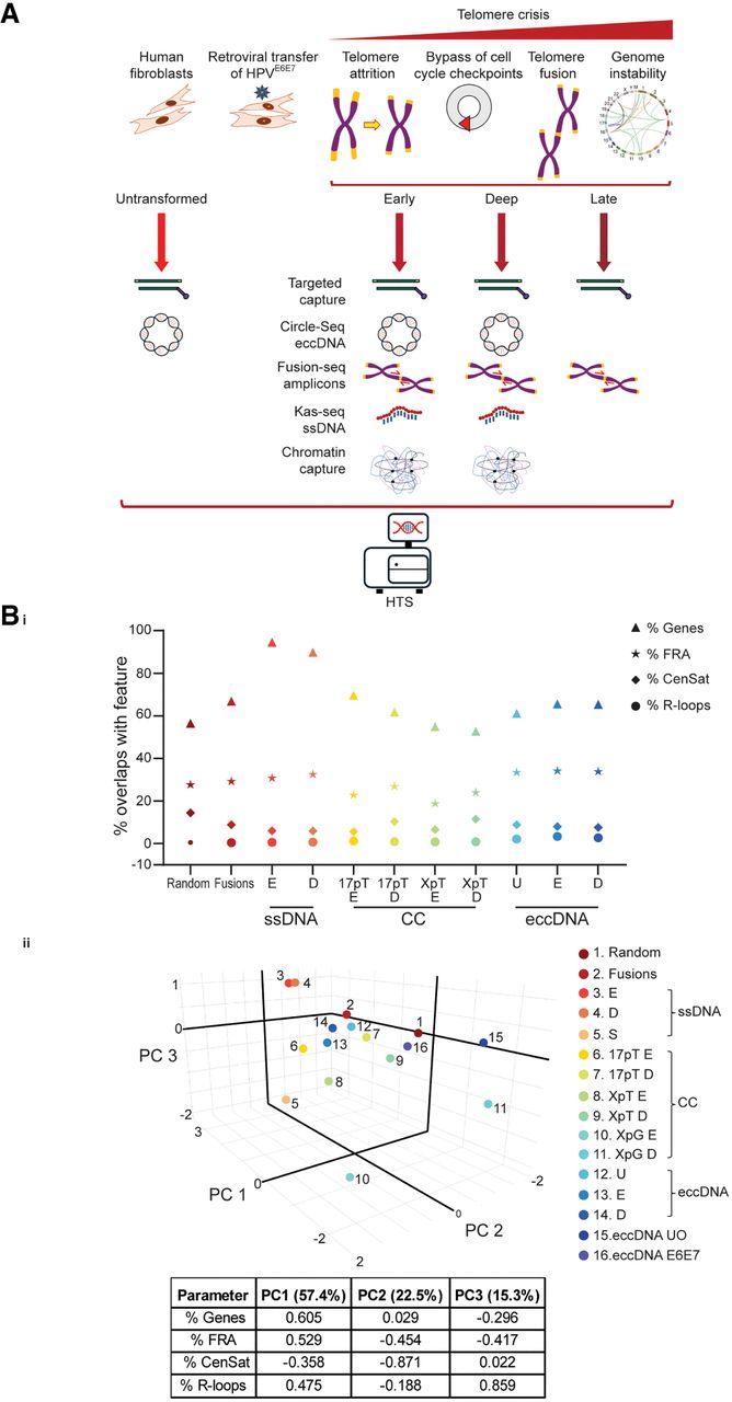
Chromatin capture (Capture-C) (Downes et al. 2022, 2023) was performed using probes specific for Chr17p (17pT) and ChrXpYp (XpT) telomere-adjacent sequences, as well as an intergenic internal ChrXpYp (XpG) control locus (Supplemental Fig. S2) to characterize the diversification of telomere contacts in crisis. Circle-Seq (Supplemental Fig. S3; Møller 2020) was employed to distinguish eccDNA produced in “normal” compared with transformed cells and determine interrelationships with telomere fusions and gross karyotypic changes. KAS-seq (Lyu et al. 2022) was used to colocalize sites of DNA replication, transcription, and repair with SVs identified through parallel sequencing processes. Targeted capture experiments were conducted across the whole range of MRC5 samples to explore potentially unstable sequences under the pressure of crisis and their associations with telomere dysfunction.
These related data sets were analyzed for recurrent locations and features informative of the origins and effectors of genomic variation (Fig. 1Bi). Sequence context proved a potent classifier, resulting in clear sample segregation by crisis stage and underlying vulnerabilities (Fig. 1Bii; Supplemental Fig. S4). Specifically, telomere fusions clustered with eccDNA and Late-crisis Capture-C data sets, suggesting common mechanisms driving genomic rearrangements.
Single-strand DNA peaks outline transcriptional networks and crisis-induced replication stress
Investigating the impact of DNA damage (KAS-seq) for chromatin remodeling, we realized a correspondence between single-strand DNA (ssDNA) signal peaks and coding sequence (Fig. 2Ai) irrespective of crisis stage. Using our crisis-cell RNA-seq data (Liddiard et al. 2021), we were able to corroborate the active expression of these genes within MRC5E6E7 cells. Compared with a data set derived from 1 million random genomic coordinates (generated using BEDTools RandomBED) (Quinlan and Hall 2010; Galaxy Community 2024), ssDNA peaks called from discrete crisis samples exhibited substantially higher proportions of genomic intervals overlapping with genes and expressed genes (Fig. 2Aii). Only the ssDNA peaks detected throughout crisis (“Shared” between data sets) displayed significant association with R-loops (Supplemental Fig. S4), suggesting that, although unique ssDNA peaks demarcate stage-specific transcription, common peaks likely represent sites of replication stress or transcription–replication clashes. Whereas the Early-crisis ssDNA peaks data set (2455 genes) was enriched for genes involved in biogenesis and proliferation (Supplemental Fig. S5A), the Deep-crisis data set (2001 genes) showed the inflammation that accompanies persistent DNA damage and cellular stress (Nassour et al. 2019; Lex et al. 2020; Dust et al. 2022; Gulen et al. 2023). Genes bearing ssDNA marks at both crisis stages (Shared; 1668 genes) were typically longer genes (Supplemental Fig. S5B) more predisposed to replication errors (Wei et al. 2016; Berkemeier et al. 2025). Global transcription declined with crisis transition (Supplemental Fig. S5C), but this was insignificant for genes closer to the telomeres with lower mean lengths (Supplemental Fig. S5D). Consonantly, ssDNA peaks in Deep crisis marked shorter genes (Supplemental Fig. S5B) closer to the chromosome termini (Supplemental Fig. S5E), affirming telomere-proximal gene expression accompanying telomere attrition (Baur et al. 2001).
Correspondence of single-strand DNA (ssDNA) peaks with transcription and telomere-interacting sites in replicative crisis. (A, i) UpSet plot (Lex et al. 2014) depicting the overlaps (intersections) between MRC5E6E7 KAS-seq data sets and coding sequence (Genes) or genes expressed by MRC5E6E7 during crisis (Expressed) (Liddiard et al. 2021). (Early, Deep) Crisis stages, (Shared) ssDNA peaks common to both stages. Connected dots indicate the data sets included in each intersection, with vertical bars showing the number of elements in that intersection, enumerated above the bars. Key intersections are boxed in red. The data set (Set) sizes in nucleotide base pairs are depicted as horizontal bars. (ii) The proportions of genomic intervals associated with the specified Kas-seq ssDNA data sets or 1 million random genomic loci that intersect with genomic features are displayed. (Early, Deep) Crisis stages, (Shared) ssDNA peaks common to both stages, (Genes) coding sequence, (Expressed) genes expressed by MRC5E6E7 during crisis (Liddiard et al. 2021), and (R-loops) three-stranded nucleic acid structures. Differences were evaluated using the N − 1 χ2 method (P-values indicated). (B) Pie charts representing the fractions of Early- and Deep-crisis genes with ssDNA enriched at 5′ or 3′ locations or detected at both 5′ and 3′ ends or throughout the entire gene body. (Undetermined) Negligible ssDNA signal. The results of N − 1 χ2 comparisons of the 5′ and 3′ ssDNA gene enrichments are denoted next to the Deep-crisis chart. (C) The 5 kb genomic sequence extending from the 3′ termini of genes with prevailing 3′ ssDNA signal was assessed for presence of putative non-B DNA repeat motifs (x-axis; defined by Advanced Biomedical Computational Science [ABCS]) (Cer et al. 2012). Incidence rates (number of motifs per kilobase of input DNA) were compared for Early- and Deep-crisis samples, and the P-value for the incidence rate ratio is indicated. (G4) G-quadruplex repeats.
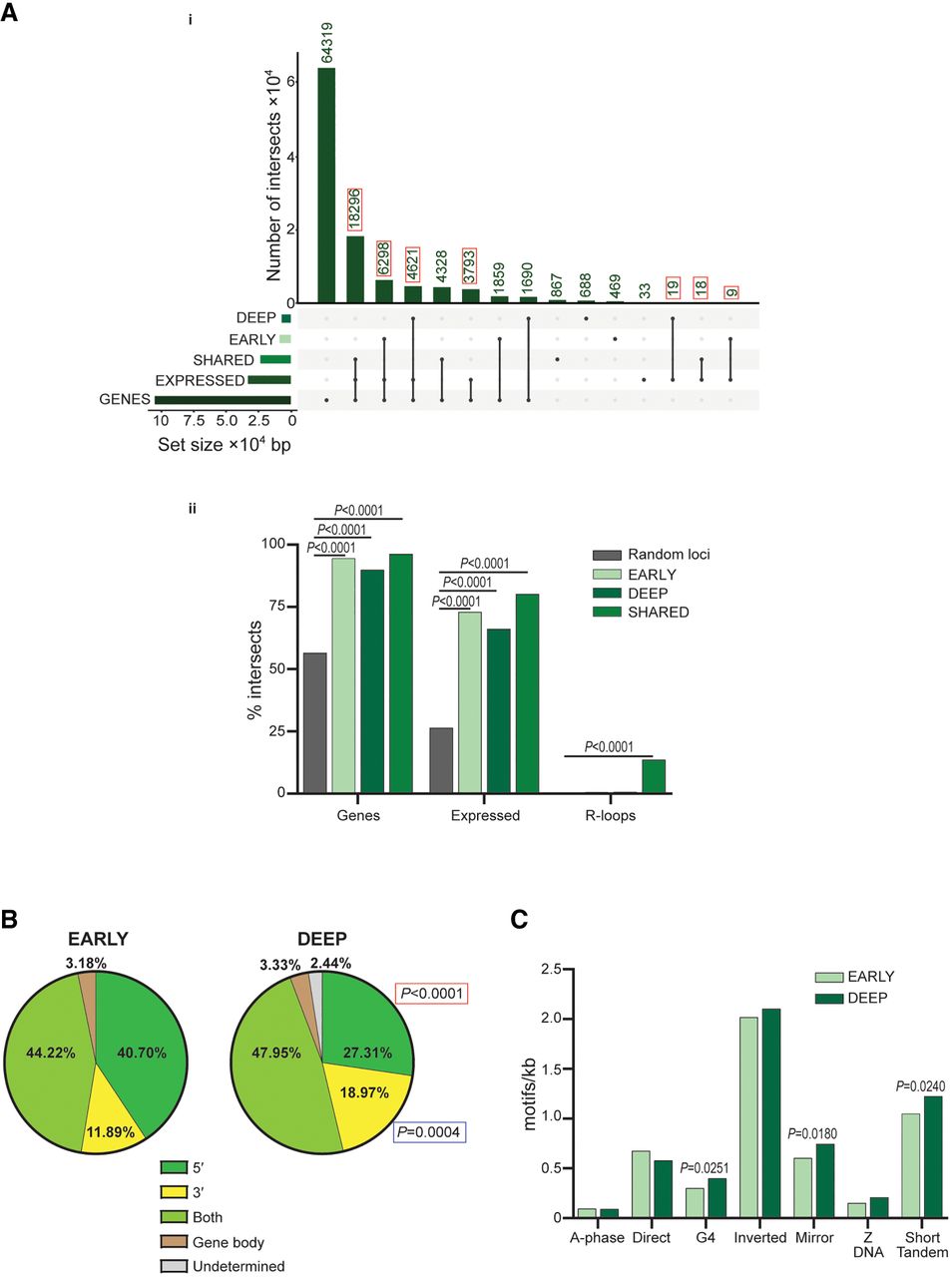
Ascribing KAS-seq signal peaks to 5′ or 3′ gene locations revealed a compelling reduction in ssDNA peaks overlapping transcription start sites (TSSs) in Deep crisis (Fig. 2B) and corresponding elevation in 3′ DNA damage characteristic of replication fork stalling and structural instability (Li and Manley 2005; Gadgil et al. 2020, 2024; Stoy et al. 2023). We observed increased incidence of G4-quadruplex, mirror, and short tandem repeat (STR) motifs downstream from genes with 3′ ssDNA peaks in Deep crisis (Fig. 2C), proposing these structures as conducive to DNA lesions and replication stress in crisis (Makova and Weissensteiner 2023; Gadgil et al. 2024). Thus, KAS-seq signal peaks may be adopted as surrogate markers of both active transcription and potential sources of replication fork stalling in replicative crisis.
Crisis reshapes the three-dimensional genome
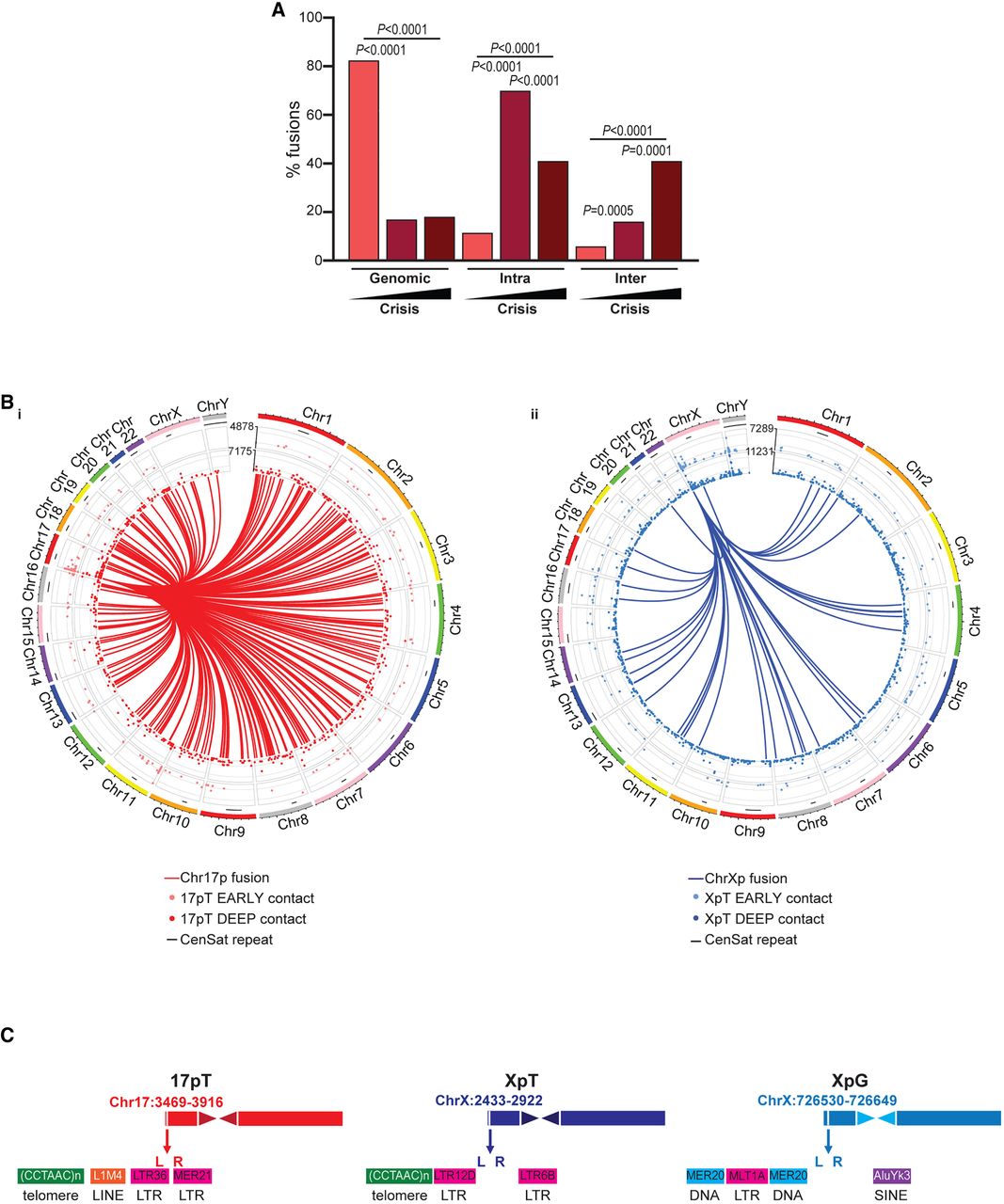
Telomere fusions amplified from MRC5E6E7 displayed progressively increasing proportions of longer-range inter- than intrachromosomal telomere–telomere rearrangements (Fig. 3A) with crisis transition. Conversely, the incidence of telomere–telomere (both intra- and inter-) fusions in Early-crisis samples was far lower than in stochastic genomic rearrangements, likely reflecting the more limited reservoir of eroded and fusogenic telomeres at this stage. Telomere-genomic recombinations may occur in the absence of telomere erosion through synapsis of DSBs in both locations. To understand the differential interactions of functional and eroded telomeres and their potential contributions to malignant recombination (Fig. 3B; Supplemental Fig. S6A,B), we performed Capture-C in Early- and Deep-crisis MRC5E6E7 using Chr17p (17pT) and ChrXp (XpT) telomere-adjacent capture probes and a nontelomeric control probe (XpG) (Fig. 3C; Supplemental Fig. S2).
The changing profiles of telomere interactions and fusions during crisis transition. (A) The proportions of mapped telomere fusion sequences generated from Early (PD23) MRC5E6E7 crisis cells in this study that were classified as “genomic” (telomere fused to internal genomic locus), “intra” (intrachromosomal; telomeres of the same chromosome fused together) or “inter” (interchromosomal; telomeres of different chromosome fused together) are presented alongside comparable data for PD45 and PD49 MRC5E6E7crisis cells detailed in our former study (Liddiard et al. 2021). The black triangle and darker colored bars indicate the progression of replicative crisis. Statistical analyses employed the N − 1 χ2 method. (B) Circos plots (Yu et al. 2018) depicting all MRC5E6E7 Early- and Deep-crisis chromatin interactions with Chr17p (17pT; i) or ChrXp (XpT; ii) telomere-adjacent probes (17pT) determined using Capture-C presented as concentric perimeter tracks. Track height indicates signal intensity. Fusions between the Chr17p (i; red) or ChrXp (ii; blue) telomeres and genomic sites span the circle centers. Peripheral black lines underneath the chromosome identities demarcate the centromere and pericentromeric (CenSat) repeats. (C) Cartoons depicting the sequence contexts and human T2T-CHM13/hs1 genomic reference (Nurk et al. 2022) coordinates of the Chr17p (17pT), ChrXp (XpT), and internal genomic control (XpG) probes used in the Capture-C experiments. L and R denote the positions of the dual oligonucleotide probes at each location (for details, see Supplemental Table S1). Adjacent DNA repeats are represented by colored blocks. (LTR) Long terminal repeat, (LINE) long interspersed repeat, (SINE) short interspersed repeat, and (DNA) DNA repeat.
We observed robust elevations in long-range (trans) interactions (Fig. 4Ai) from both telomere-adjacent (17pT and XpT) and genomic (XpG) capture probes (but not unrelated capture probes targeting five distinct genes) (Supplemental Fig. S6C) with advancing crisis. This cis-to-trans shift was accentuated by a pronounced increase in centromeric contacts (Fig. 4Aii,iii) and a complementary suppression of telomere-proximal connections (Fig. 4Aiv), potentially linked to telomere shortening (Supplemental Fig. S1; Baur et al. 2001).
Crisis represents a transition in chromatin state and long-range chromosomal interactions. (A, i) Normalized read counts for all long-range trans compared with local cis chromatin Capture-C interactions determined by the Capcruncher pipeline. Triplicate samples of the Early- and Deep-crisis MRC5E6E7 sampling points using the Chr17p and ChrXp telomere-adjacent (17pT; XpT) and the ChrXp internal genomic (XpG) probes are displayed as means with 95% confidence intervals (CIs), analyzed using paired parametric t-tests. The mean sample percentages of interactions captured by the 17pT, XpT, and XpG probes in Early and Deep crisis that overlap with ALR/alpha satellite repeats (ii), are within 10 Mb of centromere (iii), or are within 10 Mb of telomere sequences (iv) are presented and compared using the N − 1 χ2 method. (B) The proportions of unique genes identified within crisis MRC5E6E7 telomere fusions sequenced in this and our former study (Liddiard et al. 2021) that overlap with genes captured by 17pT, XpT, or XpG probes in chromatin Capture-C experiments are shown and evaluated using the N − 1 χ2 method. (C) The proportions of genomic intervals associated with telomere fusion and chromatin Capture-C data sets that intersect with genes expressed in comparable crisis stage MRC5E6E7 samples (Liddiard et al. 2021) were computed using BEDTools intersect intervals (Quinlan and Hall 2010). Intersections with MRC5E6E7 telomere fusions were compared with intersections of each individual 17pT, XpT, or XpG probe Capture-C data set using the N − 1 χ2 method. (E) Early crisis, (D) Deep crisis. (D) The proportions of genomic intervals derived from chromatin Capture-C data sets (17pT, XpT, and XpG probes; Early- and Deep-crisis stages) that intersect with DNA repeats defined by RepeatMasker (x-axis) (Smit et al. 2013) are presented. The order of the data sets displayed corresponds to the adjacent key. (DNA) DNA repeat, (LINE) long interspersed repeat, (SINE) short interspersed repeat, and (LTR) long terminal repeat. Evaluation of differences employed the N − 1 χ2 method. (E) XpT chromatin Capture-C interactions were categorized according to the presence or absence of single-nucleotide variants (SNVs) characteristic of the long (A) or short (B) ChrXp telomere allele (Baird et al. 2003). The percentages of “all” and “intra”- or “inter”-chromosomal XpT interactions are compared for the Early- and Deep-crisis stages using the N − 1 χ2 method. (F) The proportions of genomic intervals associated with ChrXp A or B total allelic contacts that intersect with genes, expressed genes, and centromeric satellite (CenSat) repeats (i) or SINE and satellite DNA repeats (ii; delineated by RepeatMasker) (Smit et al. 2013) were analyzed using the same methodology. (G) The lengths of all genes (log10 scaling; means annotated within boxes below the x-axis) captured by the 17pT and XpT subtelomere probes at Early- and Deep-crisis stages (i) and the XpT A or B alleles at both crisis stages (ii). Means with 95% CI were compared using unpaired nonparametric Mann–Whitney U tests.
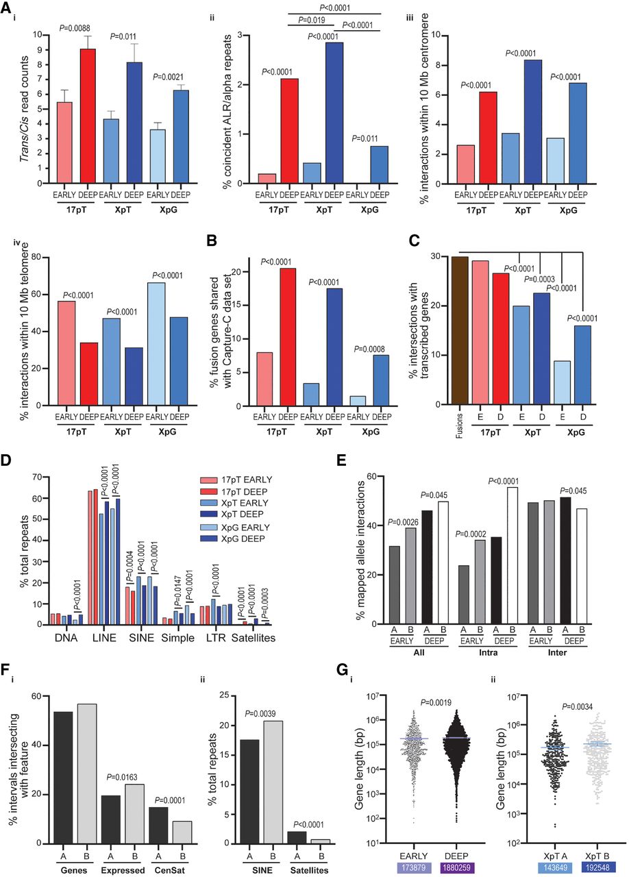
Subsets of genes interacting with telomere-adjacent probes were conjointly detected as telomere fusions in crisis MRC5E6E7 (Fig. 4B), suggesting recombination is, in part, related to the probability of stochastic contact between distant sites. Gene overlaps were functionally enriched in transmembrane signaling networks (Supplemental Fig. S6D) critical to the DNA damage response (DDR) (Maliszewska-Olejniczak and Bednarczyk 2024), indicating that genomic rearrangements incorporate genes actively expressed and required for crisis-cell viability. This creates a potential liability, whereby ongoing expression and reliance on these pathways increase the likelihood that further rearrangement or mutation compromises essential cellular processes. Intersections between Capture-C data sets and genes expressed by crisis MRC5E6E7 cells were consistently lower than for our collection of MRC5E6E7 telomere fusions but exhibited marked probe-dependent effects (Fig. 4C). Thus, transcription is less explicitly associated with subtelomere interactions than with fusions. Underlying associations with DNA repeat classes revealed only a dominance of LINE repeats at captured sites, with reductions in SINE and satellite sequences observed for all probes (Fig. 4D) in Deep crisis. Reassuringly, we detected no spurious enrichment of LTR sequences despite the prominence of these repeats at the chromosome ends (Fig. 3C).
Exploring the totality of Capture-C gene contacts, we found Deep-crisis subtelomere interactions to be specifically enriched in sodium transport genes (Supplemental Fig. S7A), and so we queried the ICGC/TCGA pancancer database (ICGC/TCGA Pan-Cancer Analysis of Whole Genomes Consortium 2020) for cognate gene abnormalities in cancer patients. We discovered aberrations in 70% patients (1808 of 2583 patients sampled), with amplifications being the most prevalent (Supplemental Fig. S7B). Variations in six of these genes (Supplemental Fig. S7C) had significant impacts for patient survival, linking the altered chromatin configurations we observed during crisis transition with cancer progression.
MRC5 cells have ChrXpYp alleles with disparate TLs (Supplemental Fig. S1C), enabling the longer (A) and shorter (B) alleles to be distinguished through heterozygous single-nucleotide polymorphisms in the telomere-adjacent DNA (Baird et al. 1995). This allelic asymmetry allows TL-dependent effects on long-range subtelomeric interactions to be assessed independently of crisis stage or replicative age, as both long and short telomere alleles can be simultaneously tracked within the same crisis cells. The shorter telomere (B) allele contributed to the greater proportion of intrachromosomal interactions throughout crisis compared with the longer telomere (A) allele (Fig. 4E), whereas no differential interchromosomal association was discerned. Both alleles echoed the diminished telomere-proximal contacts in Deep crisis (Supplemental Fig. S8A) observed with the wider data sets (Fig. 4Aiv) but only a robust association between the long allele and centromeres at the same stage (Supplemental Fig. S8B). Overall, the shorter (B) telomere allele displayed elevated intersections with expressed genes (Fig. 4Fi) and SINE repeats (Fig. 4Fii; Supplemental Fig. S8C) in contrast to the longer (A) telomere allele that had proportionally more interactions with centromere satellite (CenSat) and satellite repeats. These observations infer compartmentalization of extended repeat tracts within the nucleus, abrogated by telomere shortening that confers extraordinary mobility (Cho et al. 2014) and capacitates fusion with sister chromatids and genomic DSB (Liddiard et al. 2016). The increased mean lengths of genes captured in Deep crisis (Fig. 4Gi) or with the shorter B allele (Fig. 4Gii) is consistent with longer genes precipitating replication stress (Wei et al. 2016) and DSBs that promote cosegregation with deprotected telomeres. These results also corroborate recent findings of allele-specific chromatin organization (Irastorza-Azcarate et al. 2025).
We performed XSTREME motif discovery and enrichment analysis (Bailey 2021) in sequences flanking Capture-C probe interactions with genes also disrupted by telomere fusions (Fig. 4C) to identify organizing factors (Supplemental Fig. S9). Binding sites for chromatin insulator loop factors (including ZNF770, ZNF335, ZFX, and PITX2) (Trieu et al. 2020) were among the most enriched motifs, pinpointing topological boundaries as sites of particular vulnerability in the evolving crisis genome. Binding motifs for the inflammatory modulators BCL6 (Sawant et al. 2012) and EGR3 (Baron et al. 2015; Ogbe et al. 2015) were exclusively enriched in sequence overlaps with the telomere-adjacent capture probes and not with XpG or coding sequence probes. RNA-seq and KAS-seq data confirmed expression of BCL6 and EGR3 genes in MRC5E6E7 cells, propounding crisis-activated transcription (and the consequential disruption of this) as being instrumental in stress-induced genome reorganization (Dimitrova et al. 2008; Baron et al. 2015; Cardenas et al. 2017; Arnould et al. 2023).
Cellular stress drives a shift in eccDNA profiles
Telomere fragments (Lovejoy et al. 2012; Geiller et al. 2022) and telomere fusions (Muyas et al. 2024) have been detected as circular DNA species, and so, we next sought to clarify the interrelationship between telomere dysfunction and these specialized SVs. To ascertain the hierarchy and salient features of events (Tatman and Black 2022), we first purified, amplified, and sequenced eccDNA (Møller et al. 2016; Møller 2020; Chen et al. 2022) from Early- and Deep-crisis MRC5E6E7 fibroblasts, as well as the parental Untransformed cells (Supplemental Fig. S3). The chromosomal locations of all eccDNA junctions sequenced in four independent experiments are displayed in Figure 5A and Supplemental Figure S10A and are enumerated in Supplemental Table S1. Contrary to the high CN DNA circles serving as oncogenic drivers of specific cancers (Schwab et al. 1983; Sauter et al. 1996), we did not identify breakpoints common to replica samples (Yi et al. 2022), advocating transient and labile eccDNA formation resemblant of the unique telomere fusions that arise during crisis (Liddiard et al. 2016). Furthermore, the eccDNA we sequenced contained both coding and intergenic sequences, rather than entire genes, and varied in length from hundreds to millions of base pairs, affirming the capture of diverse eccDNA species (Yang et al. 2022).
Distinct quantities and characteristics of extrachromosomal circular DNA are detected in untransformed and crisis MRC5 cells. (A) The quantities of all sequenced eccDNA junctions amplified from Untransformed MRC5 (U; six replicas) and Early (nine replicas) and Deep (six replicas) crisis MRC5E6E7 cells are displayed by chromosome. (B) Bar chart displaying mean sizes in base pairs with 95% CI for Illumina short-read sequenced (i) or Pacific Biosciences (PacBio) long-read sequenced (ii) simple eccDNA. Mean values × 105 bp are displayed in colored boxes below the bars. Statistical evaluation was by Mann–Whitney unpaired nonparametric U tests. (C) Stacked chart displaying the relative proportions of distinct structural variants (SVs) called from the PacBio long-read eccDNA sequencing data for Untransformed MRC5 and Early- and Deep-crisis MRC5E6E7 cells. (DEL) Deletions, (DUP) duplications, (INS) insertions, (INV) inversions, (TRA) translocations, and (BND) break-end rearrangements that cannot unambiguously be categorized as a single canonical SV. Vertical black lines connect data sets evaluated using the N − 1 χ2 method, with results displayed as (*) P < 0.05, (**) P < 0.01, (***) P < 0.001, (****) P < 0.0001. (D, i) Venn diagram showing the numbers of direct gene overlaps among the eccDNA derived from Untransformed MRC5 and Early- or Deep-crisis MRC5E6E7 cells. (ii) The lengths of genes (means annotated below the x-axis) uniquely identified within genomic intervals assigned to eccDNA from Untransformed (U only) or transformed (E6E7 only) MRC5 cells are displayed in a bar chart with means and 95% CI. Mean values × 105 bp are displayed in colored boxes below the bars. Differences were assessed by unpaired parametric t-tests with Welch's correction for unequal standard deviations (SD). (E, i) The proportions of eccDNA junctions amplified from Untransformed and Early- and Deep-crisis MRC5 samples that comprise DNA segments derived from the same strand (“Simple”) or opposing strands (“Template-switch”) or distant chromosome locations (“Translocation”) are depicted as a bar chart of means with 95% CI and compared using parametric unpaired t-tests with Welch's correction. A schematic depicting DNA orientation at the eccDNA junctions (ChrA, ChrB) is shown below the chart. (ii) Simple and template-switch junctions sequenced from all samples were examined for microhomology usage >1 bp (MH; pale gray) and inserted DNA sequences >1 bp (INS; black) or blunt joins (BLUNT; white). The proportions of junctions displaying each feature are presented in a bar chart and evaluated using unpaired nonparametric Mann–Whitney U tests. (F, i) Genomic intervals for eccDNA derived from Untransformed MRC5, Early- and Deep-crisis MRC5E6E7, or subsamples comprising Untransformed-only (U only) or transformed-only (E6E7 only) were intersected with the features indicated on the x-axis. (Genes) All coding sequence, (Expressed) genes expressed in crisis MRC5E6E7, (FRA) fragile sites, (CenSat) peri/centromeric repeats, and (R-loops) trinucleotide DNA structures. Pairwise comparisons were performed using the N − 1 χ2 method and results are displayed as (*) P < 0.05, (**) P < 0.01, (***) P < 0.001, (****) P < 0.0001. (ii) The same eccDNA sample (indicated on the x-axis) genomic intervals were intersected with DNA repeats (defined by RepeatMasker) (Smit et al. 2013), and the proportions of each repeat class (right-hand color key) are displayed in a stacked bar chart. The notable differences between the discrete Untransformed-only (U only) and transformed-only (E6E7 only) were evaluated with the N − 1 χ2 method, as in i. (RC) Rolling circle repeats, (RNA) grouped representation of all classes of RNA repeats: rRNA, tRNA, snRNA, scRNA, and srpRNA.
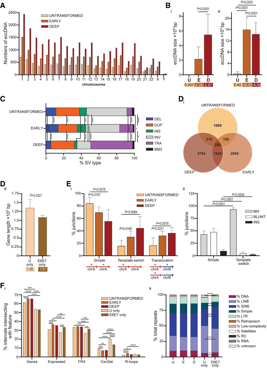
Consonant with the increased abundance of eccDNA amplified from MRC5 progressing deeper into replicative crisis (Supplemental Fig. S3Biii), we measured escalations in both mean eccDNA sizes (Fig. 5B) and complexity (Fig. 5C). Long-read sequencing revealed a substantial expansion in the proportion of eccDNA comprising DNA translocations for the Deep-crisis samples that mirrors the increasing disorganization of the crisis genome (Cleal et al. 2019) and the enhanced capacity for long-range interactions (Fig. 4A). Thus, eccDNA derived from Untransformed cells are evidently distinct from those detected in transformed MRC5E6E7 cells. Indeed, intersections across data sets demonstrated that both the overall eccDNA profiles (Supplemental Fig. S10B) and genes captured within eccDNA (Fig. 5Di) showed greater concordance between Early and Deep crisis than between Untransformed samples and either crisis stage.
Although eccDNA were smaller in Untransformed cells than in crisis cells (Fig. 5B), the genes within the genomic intervals from which they originated had longer mean lengths (Fig. 5Dii), suggesting that intermediates of late replication may circularize during normal cell division (Møller et al. 2018; Berkemeier et al. 2025; Eugen-Olsen et al. 2025). Functional enrichments in related gene networks (cell junctions and transmembrane signaling) were determined for both direct overlaps (Fig. 5Di; Supplemental Fig. S11A) and shared genomic intervals (Supplemental Figs. S10B, S11B), with crisis sample genes also reflecting metabolic divergence. Together, these observations suggest that eccDNA-associated gene content reflects cellular processes engaged during crisis, including changes in energetic demand (Turner et al. 2017).
Because eccDNA may be generated by homology-dependent and non-homology-dependent DNA repair (Cohen et al. 2006; Gadgil et al. 2024; Kang et al. 2025), we investigated eccDNA junction sequences for mechanistic insights. A prominent transformation in junction attributes was evident (Fig. 5Ei), with progressive increases in the proportions of both template-switch (opposite strand orientation) junctions and translocations, aligned with the expansion in translocations observed for the crisis compared with Untransformed samples (Fig. 5C). Although template-switched eccDNA junctions almost exclusively presented sequence microhomologies (MH) (Fig. 5Eii; Tatman and Black 2022), simple (same orientation) events were represented by blunt junctions as well as those bearing insertions or MH. These observations may signify a prevalence of replicative- or synthesis-dependent recombination processes underpinning template-switched events over larger genomic distances in contrast to nonhomologous end-joining (NHEJ) localized at simple junctions (Lee et al. 2007; Sotiriou et al. 2016; Zhu et al. 2017; Kishkevich et al. 2022; Yang et al. 2023; Hu et al. 2024; Qin et al. 2025). Thus, eccDNA provide a molecular readout of the deranged crisis genome.
Intersections of eccDNA genomic origins with sequence features (Fig. 5F; Supplemental Fig. S12) clearly distinguished events amplified from Untransformed MRC5 cells. In particular, the genomic intervals exclusive to Untransformed eccDNA (“U only”) commanded greater association with centromeric (Fig. 5Fi) and satellite, simple, and LINE DNA repeats (Fig. 5Fii; Supplemental Fig. S12A) and fewer intersections with expressed genes and SINE repeats. Conversely, crisis-derived eccDNA more commonly originated in coding sequence (including fragile sites) and SINE DNA repeats, an attribute shared with telomere fusions (Supplemental Fig. S12A). Associations with DNA repeats proved an effective means of sample stratification (Supplemental Fig. S12B), effectively separating crisis from Untransformed samples, with comparable variance contributions from each DNA repeat class.
Overall, these discoveries support divergent mechanisms for synthesis of eccDNA in Untransformed (normal) and transformed (crisis) cells. Untransformed cells exhibit low levels of small eccDNA derived from long genes and repeat tracks, likely reflecting end-joining-mediated circularization of replication byproducts. Crisis marks an upsurge in eccDNA formation, conceivably mediated by both replicative and homology-independent DNA repair processes and more often occurring within actively transcribed chromatin.
eccDNA signatures of cellular stress and malignancy
To assess the relevance of our observations in the MRC5E6E7 crisis model to human disease, we investigated the impact of environmental stress (Supplemental Fig. S13) and established malignancy (glioma) (Supplemental Fig. S14) for eccDNA profiles. Hypoxia influences both transcription (Johnson et al. 2008) and replication stress (Kindrick and Mole 2020; Ma et al. 2023), so we first explored the effects of transient (24 h) exposure to low (2%) compared with standard (20%) tissue culture oxygen (O2) levels on eccDNA production and telomere fusions in MRC5 cells. Low O2 resulted in higher eccDNA yields in the Untransformed cells (Supplemental Fig. S13Ai), homogenizing these to Early-crisis levels. Accordingly, we determined a substantial (33%) enlargement in mean eccDNA size under low-O2 conditions (Supplemental Fig. S13Aii), implicating cellular stress in the generation or maintenance of larger eccDNA. We also measured an increased incidence of simple eccDNA junctions (Supplemental Fig. S13Aiii) closer to the chromosome ends (Supplemental Fig. S13Aiv), and with enhanced associations with genes (Supplemental Fig. S13Bi), simple and low-complexity repeats (Supplemental Fig. S13Bii) relative to standard-O2 conditions. The limited sizes of the data sets constrained our ability to detect direct overlaps between cognate fusion and eccDNA samples (Supplemental Fig. S13Ci). Nonetheless, analysis at the gene level revealed shared amplifications across both SV types (Supplemental Fig. S13Cii). We discerned 24 genes (1.53% of 1571 total) in common between fusion and eccDNA data sets, with overlaps between the Early-crisis and low-O2-treated samples the most abundant. Examples of genes conjointly detected fused to telomeres and within eccDNA are displayed in Supplemental Figure S13D, in which VWF and CCSER1 appeared as fusions and eccDNA in Early-crisis low-O2-treated samples, TTN was a fusion and eccDNA in standard-O2-treated samples, and EXT1 was identified in fusions from both Early-crisis low- and standard-O2-treated samples and an eccDNA in Untransformed standard-O2-treated samples. These results indicate a degree of parity among genes susceptible to DSB and recombination by quite different processes. However, the lack of junction proximity or similarity precludes definitive assignment of sequential or related mechanisms at present.
Gliomas, particularly glioblastoma multiforme (GBM), display considerable intratumoral heterogeneity and therapy resistance that may be conferred by eccDNA-driven oncogene expression (Noorani et al. 2022). We obtained unfixed tumor and matched-adjacent tissue from three patients with astrocytoma (WHO grade 2/3; IDH1, ATRX, and TP53 mutated) (Louis et al. 2021) and three patients with GBM (WHO grade 4; TERT promoter mutated) (Supplemental Table S2). ATRX mutations are associated with decompressed chromatin at telomeres (Episkopou et al. 2014; Clynes et al. 2015) that is permissive of alternative lengthening of telomeres (ALT) and the production of extrachromosomal DNA containing telomere repeats (Lovejoy et al. 2012). We initially tested the capacity of eccDNA sequencing to detect such extrachromosomal telomere circles by evaluating the prevalence of telomere repeats (three contiguous hexamers) within SVs (Supplemental Fig. S14Ai). Notwithstanding intersample variability, eccDNA derived from astrocytoma patients exhibited a fourfold higher incidence of telomere repeat modules compared with the GBM patient samples, despite having similar eccDNA content overall (Supplemental Fig. S14Aii). We also detected examples of directly inverted telomere repeats in one astrocytoma patient sample that may indicate the circularization of pre-existing telomere fusions (Muyas et al. 2024). Junction analyses revealed salient disease-specific characteristics (Supplemental Fig. S14B), with evidence of enhanced MH usage in astrocytoma-derived eccDNA. Gene lengths were comparable across the samples (Supplemental Fig. S14Ci) but were located closer to the telomeres in eccDNA amplified from tumors compared with matched tissue samples (Supplemental Fig. S14Cii), even though TLs were not patently different (Supplemental Table S2).
Analyzing underlying genomic features exposed a hypoxic signature for the astrocytoma tumor samples (Goncalves et al. 2025) comparable with the low-O2-treated MRC5 cells. The astrocytoma (but not GBM) tumor samples shared more overlapping genomic intervals with the totality of the MRC5 low-O2 (2%) samples than the standard-O2 (20%) samples (Supplemental Fig. S14D). Repeat associations (Supplemental Fig. S14E) also mirrored those observed in low-O2 MRC5 samples, with increased intersections with simple, satellite, and low-complexity repeats (Yehuda et al. 2025) and reduced overlap with LINE elements. STRING functional protein interaction analyses revealed a single reactome pathway enrichment in VEGFA–VEGFR2 signaling (FDR 0.0125) (Supplemental Fig. S14Fi) characteristic of chronic hypoxia (Shweiki et al. 1992; Blouw et al. 2003; Godard et al. 2003) for the GBM tumor sample. Circularized fragments of oncogenes (CRK, SRC, ALK), DNA repair components (FANCC, FAN1, MGMT, FANCE, MLH1, PARP8, HIPK2), and immune effectors (HLA-E and C2) were detected in the GBM and astrocytoma (Supplemental Fig. S14F) samples, with potential impact for pathological progression. We additionally detected amplification of the EGFR gene in one GBM patient (Supplemental Fig. S14G) that was more extensive and exclusive than the recurrent punctate signal associated with simple repeats within the terminal intron. EGFR mutations and amplifications are key drivers of glioma (Libermann et al. 1985), and the same GBM patient was determined to have cytogenetic rearrangement of exons 14 and 15 of this gene (Supplemental Table S2). Thus, eccDNA profiles from patient samples capture key oncogenic processes driving malignancy.
Targeted evaluation of genome instability
Using a custom-designed targeted capture gene and genomic features panel, we pursued indices of genome-wide instability accompanying replicative crisis. We employed Manta (Chen et al. 2016) to call SV in pseudo-tumor-normal sequential MRC5 sample pairs (Early vs. Untransformed, Deep vs. Early, Late vs. Deep). Filtering for “break-end” (SV) calls outside of the targeted capture probe regions with junctions unique to individual samples revealed 38 translocations (Fig. 6A) linking 54 genes from remote loci. SVs were most abundant in Late-crisis samples (Supplemental Fig. S15A) and notably coincident with fragile sites (Supplemental Fig. S15B), highlighting replication stress as a potential mechanism for the formation of these nonclonal rearrangements.
Mutational shifts with crisis progression. (A) The genomic distributions of 38 structural variants (SVs) called by Manta (Chen et al. 2016) from targeted capture sequence data for Untransformed (U; pale gray) and MRC5E6E7 undergoing replicative crisis depicted as Circos plot (Yu et al. 2018) links. (E) Early crisis, (D) Deep crisis, and (L) Late crisis. Colors are from mid-gray to black. Genes incorporated into the SVs are numbered and decoded in Supplemental Table S3. The chromosome identities, centromere positions (denoted by CenSat repeats), and locations of the genome instability probes featured on the targeted capture panel are indicated. (B) The percentages of all single-nucleotide variants (SNV) called using VarDict (Lai et al. 2016) for pairwise pseudo-tumor-normal (Early crisis vs. Untransformed, Deep vs. Early crisis, and Late vs. Deep crisis) comparisons of MRC5 samples sequenced following targeted capture are displayed. Transitions (purine–purine or pyrimidine–pyrimidine) are underscored in orange (left); transversions (purine ↔ pyrimidine), in green (right). Comparisons of SNV proportions in progressive samples were performed with the N − 1 χ2 method, and results are displayed as (*) P < 0.05, (**) P < 0.01, (***) P < 0.001, and (****) P < 0.0001. (C) The proportions of all SNV or specifically C > T transitions or C > A transversions identified in pairwise pseudo-tumor-normal MRC5 comparisons that coincide with coding sequence (Genes) or centromeric satellites (CenSat) are displayed in a bar chart and compared using the N − 1 χ2 method. (D) Overlaps between all SNVs called from progressive pseudo-tumor-normal MRC5 sample pairings and specific repeat elements (x-axis) are displayed as proportions of the total overlaps with repeats and evaluated in the same way (P-values recorded); (SINE) Short interspersed repeat, (LINE) long interspersed repeat. (E, i) A parallel plot displaying the proportions of specific DNA repeat elements (listed along the top) comprising the total repeats intersections with the altered copy number (CN) data sets indicated on the right (lower gains and upper losses for each pairwise sample comparison). (All) The genomic intervals common to all pairwise CN gains or CN losses. The upper and lower boundaries of the proportions are annotated at the top and bottom of the chart, respectively. (LTR) Long terminal repeat, (rRNA) ribosomal RNA repeats. (ii) A bar chart displaying the most salient differentials (evaluated using the N − 1 χ2 method) between the genomic intervals common to CN gains or CN losses with respect to DNA repeat associations. (F) The distance to the telomere on the same chromosome arm (i) or centromere (ii) of CN gains and losses common to all pairwise sample comparisons; statistical assessments by Mann–Whitney unpaired nonparametric U tests.
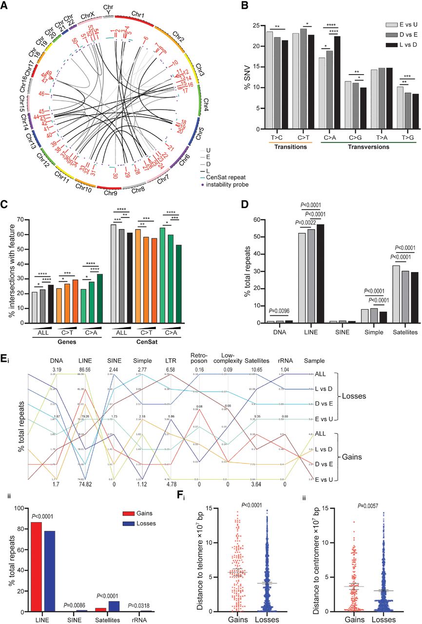
We next performed similar pairwise MRC5 sample evaluations of single-nucleotide variants (SNVs) for a more granular investigation of genome instability and mutational processes. Categorizing the nucleotide substitutions (Fig. 6B), we found that individual transitions were more common than any single transversion, but the overall proportion of transversions was greater for all samples and elevated in the latest crisis samples (Supplemental Fig. S15C). Recognizing that transversions may have more deleterious consequences compared with transitions (Freudenberg-Hua et al. 2003; Liang et al. 2021), we explored SNV that altered with crisis transit. Among all SNV classes, only C > A transversions showed a notable increase with crisis progression (Fig. 6B), as exemplified by the mutation observed within the first intron of HAVCR2 (TIM-3) (Supplemental Fig. S16A). This SNV alters putative binding sites for regulators, including FOXQ1 (HFH1) (Supplemental Fig. S16B), which, concurrently with HAVCR2, is substantially upregulated in Late-crisis MRC5E6E7 cells (Supplemental Fig. S16C), affirming the possibility of functional consequences of such SNV during replicative crisis.
In accordance with the inclusion of peri/centromeric repeat targeted probes on the custom panel, we found that >60% all identified SNVs were coincident with CenSat repeats (Fig. 6C), although this proportion declined with crisis progression, consistent with the reductions in satellite and simple repeat associations (Fig. 6D). A reciprocal rise in the proportions of SNV within coding sequence was most evident for the C > A transversions (Fig. 6C), but the overall incidence within the promoter sequence was <1% for any sample pairing, as anticipated from the panel design. These altered SNV distributions reflect a shift from replication slippage (Saayman et al. 2023; Maruta et al. 2024) or alignment ambiguity within satellite sequence (Altemose et al. 2022) toward a more compelling manifestation of the deteriorating integrity of the crisis genome revealed by our multiomic analyses (Figs. 1Figure 2.Figure 3.Figure 4.–5). This was supported by evaluating the SNV classes within their trinucleotide context (Supplemental Fig. S17), resulting in mutational signatures resembling “aging” profiles (Alexandrov et al. 2020; Degasperi et al. 2022).
Although inferring absolute CN changes from targeted capture sequence data is problematic, we were able to evaluate relative copy-number variation (CNV) using the same pseudo-tumor-normal MRC5 sample pairings for sample normalization implemented in VarScan 2 (Koboldt et al. 2012). We assessed the overlap of SNVs and CNVs with sites of ssDNA breaks and DSBs from our previous data sets (including ssDNA peaks, expressed genes, eccDNA, and telomere fusions) (Fig. 1; Liddiard et al. 2021). These genomic features effectively distinguished SNV events from CNV gains and losses in samples progressing through replicative crisis (Supplemental Fig. S18A). CNV were less often coincident with R-loops and telomere fusions than SNVs but were more frequently within genomic intervals shared with crisis-derived eccDNA, demonstrating a relationship between sporadic SVs and more enduring gross CN changes.
CN losses were more commonly associated with simple, satellite, SINE, and rRNA repeats than with CN gains (Fig. 6E), conceivably explaining the reduced incidence of SNV within these sequences during crisis (Fig. 6C,D) owing to real changes in genomic content. Pertinently, we also measured 1.38- and 1.2-fold reductions in the distance of CN losses (compared with gains) to telomere (Fig. 6Fi) or centromere (Fig. 6Fii) repeats on the same chromosome arm, respectively. This altered spatial distribution is consistent with satellite repeat rearrangements and repeat contractions reported during crisis (Stults et al. 2009; Liddiard et al. 2022), which are known to exacerbate replication stress (Gadgil et al. 2020; Shaikh et al. 2022; Ma et al. 2023) and erroneous repair (Hastings et al. 2009; Xu et al. 2017). Although extrapolation from the targeted capture methodology is limited, our data support the illustration of intrinsic instability at long genes and repeat tracts, with accumulating disruption of coding sequences as crisis advances.
Discussion
Telomere-driven replicative crisis instigates global chromatin reorganization
We previously demonstrated that inflammatory gene activation accompanies telomere fusion and genome instability in four independent human fibroblast models of replicative crisis (Liddiard et al. 2021). Here, using orthogonal sequencing approaches (Fig. 1) in an MRC5 model, we have produced an integrated characterization of replicative crisis (Supplemental Fig. S18B), sampling earlier time points after HPV16 E6E7 transduction and including Untransformed cell comparators to distinguish events driving entry into and progression through crisis.
Capture-C sequencing revealed replicative crisis to be a potent driver of expansive remodeling analogous to cellular differentiation rather than a more plastic stress response (Shanel et al. 2025). Telomere attrition reduces the density of proteins bound at the chromosome termini (Takai et al. 2010; Analikwu et al. 2025), establishing a more open chromatin configuration with crisis progression. Substantive chromatin reorganization was exemplified by the elevated long-range (trans) interactions and contacts between eroded telomeres and centromere repeats (Fig. 4A) in Deep-crisis MRC5E6E7 cells. Telomere fusions mirrored this extended engagement (Fig. 3A), with an increased prevalence of interchromosomal and concomitant reduction in local intrachromosomal recombinations with crisis progression. Allelic resolution showed preferential engagement of the shorter ChrXpYp telomere with coding sequence and intrachromosomal sites, whereas the longer telomere displayed increased associations with repeat tracks (Fig. 4F). This coalescence of the short telomere allele with expressed genes and the sister chromatid is consistent with our affirmation of this as the fusogenic allele in crisis (Liddiard et al. 2016) and the patent relationship between transcription, DSBs, and telomere fusions (Escudero et al. 2019; Liddiard et al. 2021).
Both TP53 (Scheffner et al. 1990) and RB1 (Dyson et al. 1989) function is abrogated in our MRC5E6E7 model of replicative crisis (Supplemental Fig. S1), impacting chromatin structure (Gonzalo and Blasco 2005) and cell cycle progression and DNA repair (Obanya et al. 2025). Chromatin decompaction attenuates the insulating capacity of telomeres, derepressing gene expression (Baur et al. 2001) and condensin-mediated DNA loop extrusion, thereby constraining the error-free resolution of telomere fusions (Analikwu et al. 2025) that occur with increasing frequency in crisis (Supplemental Fig. S1B). We additionally observed elevated incidence of larger and more complex eccDNA (Fig. 5) and nonclonal translocations (Fig. 6) at later stages of replicative crisis. Thus, telomere attrition and chromatin remodeling support the formation of alternative chromatin loops with extended genomic reach that enable synapsis of distant loci.
The release and redistribution around the genome (Stock et al. 2022) of DNA binding factors formerly sequestered within the telomere structure (Maillet et al. 1996; Platt et al. 2013) amplify the persistent DNA damage signaled by the eroded telomere (Supplemental Fig. S5; Cesare et al. 2013; Nassour et al. 2024). The release of chromatin fragments (including replication intermediates and eccDNA) into the cytosol (Zhang et al. 2021) and telomeric repeat-containing transcripts (TERRA) into exosomes compounds the inflammatory environment (Wang et al. 2015; Lex et al. 2020). Telomeric structural transformations further impact the transcriptome through relief from the repressive telomere position effect (TPE) (Baur et al. 2001; Rey-Millet et al. 2023) that regulates expression of critical mediators of aging and inflammation (Dong et al. 2021; Liddiard et al. 2021) enriched at the chromosome termini. Although our results distinguish telomere fusions from chromosomal interactions (Figs. 3, 4), loci represented in both data sets propose the DDR as a potent reorganizing force (Supplemental Fig. S5A; Wu et al. 2024a), with binding sites for stress response mediators including BCL6 and EGR3 being overrepresented in recombined sequences (Supplemental Fig. S9).
eccDNA profiling reveals signatures of cellular stress and disease
The relative abundance and ease of eccDNA purification from crisis cells and patient samples make these SVs more attractive and robust indicators of genome instability compared with telomere fusions, particularly when input material is scarce. Notably, eccDNA derived from transformed MRC5E6E7 cells were quantitatively and qualitatively distinct from those amplified from Untransformed MRC5 (Fig. 5; Supplemental Figs. S10–S13), corroborating their potential as surrogates of malignancy (Bruhm et al. 2025). Lower yields of smaller eccDNA were harvested from Untransformed than from transformed cells (Fig. 5), and these were typified by simple junctions originating from longer genes and repeat elements that included centromeres and other satellite sequences. These characteristics suggest a constitutive NHEJ-mediated circularization (Zhu et al. 2017; Hu et al. 2024; Kang et al. 2025; Qin et al. 2025) of the byproducts of replication and transcription at challenging repeat tracks and late-replicating genes with limited impact for cellular function (Møller et al. 2018). In contrast, eccDNA amplified from crisis MRC5E6E7 captured shorter genes with greater proportions of template-switched junctions and long-range complex translocation events, compatible with our Capture-C data and suggestive of replicative repair (Sotiriou et al. 2016; Kishkevich et al. 2022; Zhang et al. 2023). Furthermore, we detected increased association with transcribed genes and SINE repeats, linking this transition to transcriptional reprogramming induced by crisis (Liddiard et al. 2021).
Regarding eccDNA as tangible indices of genome instability, we determined a sensitivity of profiles to environmental (Supplemental Fig. S13) and pathological stressors (Supplemental Fig. S14). Under conditions of transient low O2 (2%), eccDNA purified from Untransformed MRC5 approximated the yields and size distributions characteristic of crisis MRC5E6E7 eccDNA (Supplemental Fig. S13A), independent from effects on cell viability or proliferation. Cellular responses to mild hypoxia (Hu et al. 2003; Johnson et al. 2008; Kindrick and Mole 2020; Wu et al. 2022) resulted in eccDNA enriched in simple junctions capturing genes closer to telomeres, potentially reflecting NHEJ activity within sequences transcribed from the reservoir of inflammatory and immune regulators close to the chromosome ends (Lex et al. 2020; Lopes et al. 2021). Testifying to the replication stress exacerbated by hypoxia, eccDNA from cells cultured in low-O2 conditions were enriched in simple, satellite, and low-complexity repeat motifs that may challenge replication fork or transcriptional progression (Hammond et al. 2002; Chan et al. 2008; Yehuda et al. 2025), facilitating eccDNA biogenesis (Qin et al. 2025). Among our cohort of glioma patients (Supplemental Table S2), eccDNA properties distinguished astrocytoma from GBM pathology and tumor tissue from matched-adjacent samples (Supplemental Fig. S14). ATRX-mutated astrocytomas produced eccDNA enriched for telomeric and replication-stalling repeats (Clynes et al. 2015; Nguyen et al. 2017), with extended MH at junctions, consistent with replication stress and end-joining repair (Wimberly et al. 2013; Ngo et al. 2021). Whereas the astrocytoma tumor samples displayed significant parallels with MRC5 low-O2 cultures (Supplemental Fig. S14D; Goncalves et al. 2025), the GBM tumors evidenced functional gene networks characteristic of the hypoxic environment that drives this aggressive pathology (Supplemental Fig. S14F; Park and Lee 2022; Marallano et al. 2024). Thus, by analyzing eccDNA contents and junction features, we have been able to surmise both active gene networks and sites of replication stress. In a clinical setting, eccDNA profiling could likewise identify critical cancer dependencies for novel targeting strategies and therapeutic monitoring (Li et al. 2023; Wang et al. 2025).
Interrelationship of SVs
We previously reported the assimilation of circular plasmid and mitochondrial DNA into telomere fusions (Liddiard et al. 2016), raising the possibility of eccDNA being captured by deprotected telomeres during crisis. Moreover, we have exposed the association between telomere fusions, eccDNA, and sites of CNVs (Supplemental Fig. S18; Escudero et al. 2019; Liddiard et al. 2021) symptomatic of DNA excision and extrachromosomal amplification (Von Hoff et al. 1992; Verhaak et al. 2019). Accordingly, we identified recurrent genes incorporated into telomere fusions and eccDNA amplified from parallel MRC5 cell cultures, confirming that destabilized loci are substrates for both forms of SVs (Supplemental Fig. S13D). With clinical relevance, we identified eccDNA comprising inverted telomere repeats amplified from a patient with ATRX-mutated astrocytoma, consistent with the production of telomeric circles in this cancer (Cesare and Griffith 2004). The concurrent amplification and cytogenetic rearrangement of EGFR observed for one GBM patient (Supplemental Fig. S14G) are also suggestive of successive or interdependent mutational processes. Although we did not identify shared junctions among distinct SVs in parallel analyses, our findings sustain the possibility of a common origin for telomere fusions and eccDNA in malignancy.
Overall, our integrated analyses reveal the critical contributions of telomere and centromere repeats to chromatin reorganization under conditions of replicative and environmental stress. We demonstrate the interdependence of telomere attrition, replication, and transcription in the collective translation of the cellular stress response into a three-dimensional structural variation within the evolving cancer genome (Supplemental Fig. S18B). Our data support a model of replicative crisis in which the inherent instability of long, late-replicating sequences is progressively compounded by increasingly complex long-range interactions that disrupt crisis-response genes, shaping both cancer genotype and phenotype. We additionally uncover the unrealized potential of eccDNA as biomarkers of pathological processes, even in the absence of clonal amplifications. These findings highlight novel diagnostic opportunities and provide mechanistic insights into genome instability in replicative crisis.
Methods
Cells and treatments
MRC5 cells were authenticated by STR profiling with the American Type Culture Collection (ATCC) and routinely screened for the absence of mycoplasma. Cells were cultured in Eagle's Minimum Essential Medium (EMEM) supplemented with 1× nonessential amino acids, 10% (v/v) FCS, 1 × 10⁵ IU/L penicillin, 100 mg/L streptomycin, and 2 mm glutamine and buffered with 0.2% NaHCO3 solution.
The crisis MRC5 cells were generated in our previous publication (Liddiard et al. 2021) using amphitropic retroviral vectors containing HPV16 E6 and E7 oncogenes (E6E7) and a neomycin-resistance cassette (NEO), as outlined previously (Bond et al. 1999). Selection of transduced and expressing MRC5E6E7 cells was by culture in the presence of Geneticin (G418) at 0.4 mg/mL. Cells were maintained at 70%–85% confluency, with PD calculated at each passage following cell counts using an NC-3000 image cytometer (Chemometec). The PDs indicated in this paper refer to population growth after the point of retroviral transduction because this constitutes a bottleneck event (Supplemental Fig. S1A). Crisis staging (Early, Deep, and Late) was assigned according to the appearance and accumulation of telomere fusions at multiple chromosome ends (Supplemental Fig. S1B) following telomere attrition (Supplemental Fig. S1C).
For most experiments, cells were cultured in 20% O2 and 5% CO2 at 37°C. For experiments comparing standard with low-O2 stimuli, parallel cell cultures of 7 × 10⁵ Untransformed MRC5 or Early-crisis (PD23) MRC5E6E7 were established in incubators with 20% or 2% O2, respectively, with nitrogen gas compensation and regular monitoring with a handheld gas analyzer (Geotech G100).
Cell cycle analyses were performed using the Chemometec fixed cell cycle DAPI assay and analyzed using Floreada.io software.
Chromatin capture
Capture-C experiments were performed for MRC5E6E7 Early-crisis (PD23) and Deep-crisis (PD47) cells according to the method previously described (Downes et al. 2022, 2023). NlaIII restriction enzyme (NEB R0125) cleaved nuclear 3C libraries were prepared from each of three replicas of independent cell cultures of MRC5E6E7 cells for each time point (5 × 10⁶ cells each). Evaluation of library quality was made by agarose gel electrophoresis and qPCR. Libraries were sheared to 200 bp using an ME220 Covaris focused ultrasonicator and indexed using NEBNext multiplex oligos for Illumina (primer sets 1–3; E7335S, E7500S, and E7710S) in a modification of the NEBNext Ultra II DNA library prep kit for Illumina protocol. Each library was subjected to two rounds of capture hybridization with each HPLC-purified 5′ biotinylated viewpoint probe set (Merck) (Supplemental Table S1) and amplification ahead of 150 bp paired-end sequencing on an Illumina MiSeq. The stringent design of the viewpoint probes to capture interactions from the Chr17p and ChrXpYp (abridged to ChrXp throughout) subtelomeres was facilitated by the CapSequm tool (Davies et al. 2016). A nontelomeric control probe (XpG) was also designed within a noncoding locus 724 kb 5′ of the ChrXp subtelomere (XpT) probe for comparison with the subtelomere sequence context (Fig. 2F; Supplemental Fig. S2). Agilent TapeStation analyses were performed at multiple stages to verify library yields and size profiles.
Initial data analysis steps employed the CapCruncher software pipeline (Downes et al. 2022) to call genomic loci captured by each probe and produce BAM files for visualization using the Integrative Genomics Viewer (IGV) (Robinson et al. 2011). For evaluations of long-range interchromosomal interactions, IGV was used to delineate and exclude local cis-interactions from downstream analyses. A signal threshold of 500 was applied to filter the remaining capture sites to mitigate the contribution of nonspecific interactions to the analyses. Corroboration of the credibility of these trans interactions was provided by intersections with orthogonal data sets, including telomere fusion sequencing and eccDNA sequencing. BLAT was used to verify selected alignments (Kent 2002). Allelic segregation of the ChrXp data was achieved by partitioning subtelomere alignments according to definitive presence of single-nucleotide polymorphisms diagnostic of the long (A) or short (B) allele (Baird et al. 1995).
KAS-seq ssDNA mapping
KAS-seq identification of ssDNA genomic locations was implemented according to the method of Lyu et al. (2022). Three Early-crisis (PD23) and Deep-crisis (PD47) MRC5E6E7 samples were labeled with N3-kethoxal, sheared to 200 bp with the Covaris ME220 ultrasonicator, and enriched following biotinylation using the AccuraDX KAS-direct ssDNA labeling and enrichment kit. Sequencing libraries were prepared from enriched and nonenriched fractions of the same samples (used as input controls) using xGen ssDNA low-input DNA library prep (IDT 10009859) and xGen UDI primers (IDT 10005975). Sequencing employed the Illumina NextSeq 550 high-output 75 bp single-end sequencing platform to meet the protocol requirements of 30 million reads per library. Ultimately, one crisis sample resulted in a low signal sequencing output and was eliminated from the downstream analyses.
Preliminary read processing and alignment was executed with KAS-Analyzer (V1.0) (Lyu et al. 2023), and ssDNA signal peaks in the enriched Early- and Deep-crisis samples compared with the input controls were identified using epic2 (Stovner and Sætrom 2019). DiffBind (http://bioconductor.org/packages/release/bioc/vignettes/DiffBind/inst/doc/DiffBind.pdf; Ross-Innes et al. 2012) was used to perform differential analysis of consensus peaks, distinguishing peaks unique and common to the Early- and Deep-crisis states. An evaluation of false-discovery rates (FDR) and fold changes of the differences was also reported. Full commands and settings developed for this study are available at GitHub (https://github.com/e-coral/crisis_analyses) and in the Supplemental Material.
Circle-seq
Glioma tissue samples were obtained from adult patients following informed consent, under the approval of the Welsh Neuroscience Research Tissue Bank (reference 19/WA/0058). Individual identifying information is not included in this paper. The methodology established by Møller (2020) was used to purify, amplify, and sequence eccDNA from Untransformed MRC5, MRC5E6E7, and glioma patient tissue samples (Supplemental Fig. S3). Approximately 7.5 × 10⁵ cells or 4 mm glioma tissue biopsies were harvested and processed for circular DNA extraction using the plasmid mini AX DNA kit (A&A Biotechnology). Digestion with Proteinase K was for 48 h at 700 RPM and 50°C; reduction of mitochondrial DNA (mtDNA) with MssI (PmeI) was for 1 h at 37°C; and elimination of linear genomic DNA involved five sequential doses of 25 U/sample plasmid-safe DNase (Biosearch Technologies) over 36 h at 37°C. Purified eccDNA were amplified by rolling circle amplification (RCA) with φ29 polymerase (Qiagen REPLI-g mini kit) over 60 h and were visualized by SYBR gold staining of electrophoresed products. Amplified eccDNA were sheared to 300–500 bp by ultrasonication with the Covaris M220 for Illumina 150 bp paired-end sequencing. Libraries from 500 ng sheared eccDNA were prepared utilizing the NEBNext Ultra II DNA library prep kit for Illumina and indices supplied in the NEBNext multiplex oligos for Illumina (primer sets 1–3). Agilent TapeStation analyses were performed at multiple stages to verify yields and size profiles. Samples were multiplexed and sequenced on the Illumina MiSeq.
Parallel Pacific Biosciences (PacBio) long-read sequencing experiments were also performed for Untransformed MRC5 and Early- and Deep-crisis MRC5E6E7 cells, as well as a single glioma tumor sample in pre- and post-RCA formats. To preserve long-read integrity, eccDNA samples were debranched with T7 endonuclease I for 1 h at 37°C, purified using Agencourt AMPure XP magnetic beads at a 1:1 ratio, and visualized by SYBR gold staining of electrophoresed products. Multiplexed amplicon libraries were prepared using the SMRTbell prep kit 3.0 (102-182-700) in conjunction with the SMRTbell barcoded adapter plate 3.0 (102-009-200). Verification and sizing of amplicons were conducted using an Agilent fragment analyzer system and Qubit 4 Fluorometer. One hundred nanograms of each adapted amplicon was pooled for PacBio sequencing using the Sequel II sequencing kit 2.0 (102-194-400) and SMRT cell 8m single-use tray (102-281-700). Sequencing was over 30 h, resulting in 15 Gb HiFi reads.
Targeted capture sequencing experiments
For a focused investigation of genomic instability during replicative crisis, a custom targeted capture panel was designed incorporating 68 genes recurrently identified in our SV data sets; three with full-length probe coverage, 64 with exon-only (coding and UTR with 50 bp flanks) coverage, and one intronic MSI locus. The panel additionally contained probes for capturing potentially unstable repeat elements, including selected LINE, centromeric satellites, subtelomere sequences, tRNA and rRNA clusters, G-quadruplex binding motifs, and an MSI locus (details available on request). The custom panel was synthesized and quality validated by Twist Bioscience.
The Twist Bioscience cfDNA library preparation kit was used to prepare sequencing libraries from 50 ng each of MRC5 Untransformed and MRC5E6e7 Early-crisis (PD23), Deep-crisis (PD47), and Late-crisis (PD57) genomic DNA that had been sheared (Covaris) to a mean fragment size of 350 bp. Amplified libraries were subjected to a 16 h hybridization with the custom probe panel at 70°C in a thermocycler. Following targeted enrichment, multiplexed libraries were sequenced using Illumina NextSeq 550 high-output 300 cycle (2 × 150 bp) flow cell and reagents to achieve approximately 1500× coverage of the 8 Mb panel.
Code availability
New computer code developed in this research is available at GitHub (https://github.com/e-coral) and as Supplemental Code.
Data access
All raw and curated sequencing data generated in this study have been submitted to the NCBI BioProject database (https://www.ncbi.nlm.nih.gov/bioproject/) under the accession number PRJNA1181623.
Competing interest statement
The authors declare no competing interests.
Acknowledgments
This research was funded by Cancer Research UK grant C17199/A29202. H.B. is supported by the Royal College of Surgeons (RCS) England Research fellowship (520619) and Brain Research UK PhD studentship (100044). We thank Joanne Morgan, NGS Technical Manager, Division of Psychological Medicine and Clinical Neurosciences, Cardiff University School of Medicine (PacBio sequencing). We thank the Hughes Group, Genome Biology, University of Oxford Medical Sciences Division (Capture-C experiment consultation and training); Wales Gene Park, Wales Genomic Health Centre, Cardiff (Illumina sequencing); and the Welsh Neuroscience Research Tissue Bank (glioma patient samples). Some of the results in this study are based on data generated by the TCGA Research Network (https://www.cancer.gov/tcga).
Author contributions: K.L. conceived, designed, and executed the project; analyzed the data; and prepared the manuscript for publication. E.C. and K.C. provided bioinformatics expertise, mapped all the data sets to the T2T CHM13v2.0/hs1 human reference, and devised new scripts for data comparisons. H.B. was instrumental in obtaining the glioma patient samples and also conducted telomere length measurements for these samples. D.M.B. secured the funding that allowed the project to proceed and provided critical evaluation of the manuscript.
Footnotes
[1] Supplementary material [Supplemental material is available for this article.]
[2] Article published online before print. Article, supplemental material, and publication date are at https://www.genome.org/cgi/doi/10.1101/gr.281373.125.
[3] Freely available online through the Genome Research Open Access option.
References
- ↵Alexandrov LB, Kim J, Haradhvala NJ, Huang MN, Tian Ng AW, Wu Y, Boot A, Covington KR, Gordenin DA, Bergstrom EN, 2020. The repertoire of mutational signatures in human cancer. Nature 578: 94–101. 10.1038/s41586-020-1943-3
- ↵Alitalo K, Schwab M, Lin CC, Varmus HE, Bishop JM. 1983. Homogeneously staining chromosomal regions contain amplified copies of an abundantly expressed cellular oncogene (c-myc) in malignant neuroendocrine cells from a human colon carcinoma. Proc Natl Acad Sci 80: 1707–1711. 10.1073/pnas.80.6.1707
- ↵Altemose N, Logsdon GA, Bzikadze AV, Sidhwani P, Langley SA, Caldas GV, Hoyt SJ, Uralsky L, Ryabov FD, Shew CJ, 2022. Complete genomic and epigenetic maps of human centromeres. Science 376: eabl4178. 10.1126/science.abl4178
- ↵Analikwu BT, Deshayes A, van der Torre J, Guérin TM, Katan AJ, Béneut C, Barth R, Phipps J, Scolari V, Veaute X, 2025. Telomeres stall DNA loop extrusion by condensin. Cell Rep 44: 115900. 10.1016/j.celrep.2025.115900
- ↵Arlt MF, Mulle JG, Schaibley VM, Ragland RL, Durkin SG, Warren ST, Glover TW. 2009. Replication stress induces genome-wide copy number changes in human cells that resemble polymorphic and pathogenic variants. Am J Hum Genet 84: 339–350. 10.1016/j.ajhg.2009.01.024
- ↵Arnould C, Rocher V, Saur F, Bader AS, Muzzopappa F, Collins S, Lesage E, Le Bozec B, Puget N, Clouaire T, 2023. Chromatin compartmentalization regulates the response to DNA damage. Nature 623: 183–192. 10.1038/s41586-023-06635-y
- ↵Bailey TL. 2021. STREME: accurate and versatile sequence motif discovery. Bioinformatics 37: 2834–2840. 10.1093/bioinformatics/btab203
- ↵Baird DM, Jeffreys AJ, Royle NJ. 1995. Mechanisms underlying telomere repeat turnover, revealed by hypervariable variant repeat distribution patterns in the human Xp/Yp telomere. EMBO J 14: 5433–5443. 10.1002/j.1460-2075.1995.tb00227.x
- ↵Baird DM, Rowson J, Wynford-Thomas D, Kipling D. 2003. Extensive allelic variation and ultrashort telomeres in senescent human cells. Nat Genet 33: 203–207. 10.1038/ng1084
- ↵Baron VT, Pio R, Jia Z, Mercola D. 2015. Early growth response 3 regulates genes of inflammation and directly activates IL6 and IL8 expression in prostate cancer. Br J Cancer 112: 755–764. 10.1038/bjc.2014.622
- ↵Baur JA, Zou Y, Shay JW, Wright WE. 2001. Telomere position effect in human cells. Science 292: 2075–2077. 10.1126/science.1062329
- ↵Behrouzi R, Clipson A, Simpson KL, Blackhall F, Rothwell DG, Dive C, Mouliere F. 2025. Cell-free and extrachromosomal DNA profiling of small cell lung cancer. Trends Mol Med 31: 64–78. 10.1016/j.molmed.2024.08.004
- ↵Berkemeier F, Cook PR, Boemo MA. 2025. DNA replication timing reveals genome-wide features of transcription and fragility. Nat Commun 16: 4658. 10.1038/s41467-025-59991-w
- ↵Blouw B, Song H, Tihan T, Bosze J, Ferrara N, Gerber HP, Johnson RS, Bergers G. 2003. The hypoxic response of tumors is dependent on their microenvironment. Cancer Cell 4: 133–146. 10.1016/S1535-6108(03)00194-6
- ↵Böhly N, Schmidt A-K, Zhang X, Slusarenko BO, Hennecke M, Kschischo M, Bastians H. 2022. Increased replication origin firing links replication stress to whole chromosomal instability in human cancer. Cell Rep 41: 111836. 10.1016/j.celrep.2022.111836
- ↵Bond JA, Haughton MF, Rowson JM, Smith PJ, Gire V, Wynford-Thomas D, Wyllie FS. 1999. Control of replicative life span in human cells: barriers to clonal expansion intermediate between M1 senescence and M2 crisis. Mol Cell Biol 19: 3103–3114. 10.1128/MCB.19.4.3103
- ↵Bruhm DC, Vulpescu NA, Foda ZH, Phallen J, Scharpf RB, Velculescu VE. 2025. Genomic and fragmentomic landscapes of cell-free DNA for early cancer detection. Nat Rev Cancer 25: 341–358. 10.1038/s41568-025-00795-x
- ↵Capper R, Britt-Compton B, Tankimanova M, Rowson J, Letsolo B, Man S, Haughton M, Baird DM. 2007. The nature of telomere fusion and a definition of the critical telomere length in human cells. Genes Dev 21: 2495–2508. 10.1101/gad.439107
- ↵Cardenas MG, Oswald E, Yu W, Xue F, MacKerell AD, Melnick AM. 2017. The expanding role of the BCL6 oncoprotein as a cancer therapeutic target. Clin Cancer Res 23: 885–893. 10.1158/1078-0432.CCR-16-2071
- ↵Cer RZ, Bruce KH, Donohue DE, Temiz NA, Mudunuri US, Yi M, Volfovsky N, Bacolla A, Luke BT, Collins JR, 2012. Searching for non-B DNA-forming motifs using nBMST (non-B DNA motif search tool). Curr Protoc Hum Genet Chapter 18: Unit 18.7.1–22. 10.1002/0471142905.hg1807s73
- ↵Cesare AJ, Griffith JD. 2004. Telomeric DNA in ALT cells is characterized by free telomeric circles and heterogeneous t-loops. Mol Cell Biol 24: 9948–9957. 10.1128/MCB.24.22.9948-9957.2004
- ↵Cesare AJ, Hayashi MT, Crabbe L, Karlseder J. 2013. The telomere deprotection response is functionally distinct from the genomic DNA damage response. Mol Cell 51: 141–155. 10.1016/j.molcel.2013.06.006
- ↵Chan N, Koritzinsky M, Zhao H, Bindra R, Glazer PM, Powell S, Belmaaza A, Wouters B, Bristow RG. 2008. Chronic hypoxia decreases synthesis of homologous recombination proteins to offset chemoresistance and radioresistance. Cancer Res 68: 605–614. 10.1158/0008-5472.CAN-07-5472
- ↵Chen X, Schulz-Trieglaff O, Shaw R, Barnes B, Schlesinger F, Källberg M, Cox AJ, Kruglyak S, Saunders CT. 2016. Manta: rapid detection of structural variants and indels for germline and cancer sequencing applications. Bioinformatics 32: 1220–1222. 10.1093/bioinformatics/btv710
- ↵Chen Z, Qi Y, He J, Xu C, Ge Q, Zhuo W, Si J, Chen S. 2022. Distribution and characterization of extrachromosomal circular DNA in colorectal cancer. Mol Biomed 3: 38. 10.1186/s43556-022-00104-0
- ↵Cho NW, Dilley RL, Lampson MA, Greenberg RA. 2014. Interchromosomal homology searches drive directional ALT telomere movement and synapsis. Cell 159: 108–121. 10.1016/j.cell.2014.08.030
- ↵Cleal K, Jones RE, Grimstead JW, Hendrickson EA, Baird DM. 2019. Chromothripsis during telomere crisis is independent of NHEJ, and consistent with a replicative origin. Genome Res 29: 737–749. 10.1101/gr.240705.118
- ↵Clynes D, Jelinska C, Xella B, Ayyub H, Scott C, Mitson M, Taylor S, Higgs DR, Gibbons RJ. 2015. Suppression of the alternative lengthening of telomere pathway by the chromatin remodelling factor ATRX. Nat Commun 6: 7538. 10.1038/ncomms8538
- ↵Cohen Z, Bacharach E, Lavi S. 2006. Mouse major satellite DNA is prone to eccDNA formation via DNA ligase IV-dependent pathway. Oncogene 25: 4515–4524. 10.1038/sj.onc.1209485
- ↵Counter CM, Avilion AA, LeFeuvre CE, Stewart NG, Greider CW, Harley CB, Bacchetti S. 1992. Telomere shortening associated with chromosome instability is arrested in immortal cells which express telomerase activity. EMBO J 11: 1921–1929. 10.1002/j.1460-2075.1992.tb05245.x
- ↵Davies JOJ, Telenius JM, McGowan SJ, Roberts NA, Taylor S, Higgs DR, Hughes JR. 2016. Multiplexed analysis of chromosome conformation at vastly improved sensitivity. Nat Methods 13: 74–80. 10.1038/nmeth.3664
- ↵Degasperi A, Zou X, Amarante TD, Martinez-Martinez A, Koh GCC, Dias JML, Heskin L, Chmelova L, Rinaldi G, Wang VYW, 2022. Substitution mutational signatures in whole-genome-sequenced cancers in the UK population. Science 376: science.abl9283. 10.1126/science.abl9283
- ↵Dekker J, Rippe K, Dekker M, Kleckner N. 2002. Capturing chromosome conformation. Science 295: 1306–1311. 10.1126/science.1067799
- ↵Dimitrova N, Chen Y-CM, Spector DL, de Lange T. 2008. 53BP1 promotes non-homologous end joining of telomeres by increasing chromatin mobility. Nature 456: 524–528. 10.1038/nature07433
- ↵Dixon JR, Selvaraj S, Yue F, Kim A, Li Y, Shen Y, Hu M, Liu JS, Ren B. 2012. Topological domains in mammalian genomes identified by analysis of chromatin interactions. Nature 485: 376–380. 10.1038/nature11082
- ↵Dong X, Sun S, Zhang L, Kim S, Tu Z, Montagna C, Maslov AY, Suh Y, Wang T, Campisi J, 2021. Age-related telomere attrition causes aberrant gene expression in sub-telomeric regions. Aging Cell 20: e13357. 10.1111/acel.13357
- ↵Downes DJ, Smith AL, Karpinska MA, Velychko T, Rue-Albrecht K, Sims D, Milne TA, Davies JOJ, Oudelaar AM, Hughes JR. 2022. Capture-C: a modular and flexible approach for high-resolution chromosome conformation capture. Nat Protoc 17: 445–475. 10.1038/s41596-021-00651-w
- ↵Downes DJ, Smith AL, Karpinska MA, Velychko T, Rue-Albrecht K, Sims D, Milne TA, Davies JOJ, Oudelaar AM, Hughes JR. 2023. Author correction: Capture-C: a modular and flexible approach for high-resolution chromosome conformation capture. Nat Protoc 18: 3155. 10.1038/s41596-023-00860-5
- ↵Dubois F, Sidiropoulos N, Weischenfeldt J, Beroukhim R. 2022. Structural variations in cancer and the 3D genome. Nat Rev Cancer 22: 533–546. 10.1038/s41568-022-00488-9
- ↵Dust K, Carpenter M, Chen JC-y, Grant C, McCorrister S, Westmacott GR, Severini A. 2022. Human papillomavirus 16 E6 and E7 oncoproteins alter the abundance of proteins associated with DNA damage response, immune signaling and epidermal differentiation. Viruses 14: 1764. 10.3390/v14081764
- ↵Dyson N, Howley PM, Münger K, Harlow E. 1989. The human papilloma virus-16 E7 oncoprotein is able to bind to the retinoblastoma gene product. Science 243: 934–937. 10.1126/science.2537532
- ↵Episkopou H, Draskovic I, Van Beneden A, Tilman G, Mattiussi M, Gobin M, Arnoult N, Londoño-Vallejo A, Decottignies A. 2014. Alternative lengthening of telomeres is characterized by reduced compaction of telomeric chromatin. Nucleic Acids Res 42: 4391–4405. 10.1093/nar/gku114
- ↵Escudero L, Cleal K, Ashelford K, Fegan C, Pepper C, Liddiard K, Baird DM. 2019. Telomere fusions associate with coding sequence and copy number alterations in CLL. Leukemia 33: 2093–2097. 10.1038/s41375-019-0423-y
- ↵Eugen-Olsen RAB, Hariprakash JM, Oestergaard VH, Regenberg B. 2025. Molecular mechanisms of extrachromosomal circular DNA formation. Nucleic Acids Res 53: gkaf122. 10.1093/nar/gkaf122
- ↵Fong YW, Cattoglio C, Tjian R. 2013. The intertwined roles of transcription and repair proteins. Mol Cell 52: 291–302. 10.1016/j.molcel.2013.10.018
- ↵Freudenberg-Hua Y, Freudenberg J, Kluck N, Cichon S, Propping P, Nöthen MM. 2003. Single nucleotide variation analysis in 65 candidate genes for CNS disorders in a representative sample of the European population. Genome Res 13: 2271–2276. 10.1101/gr.1299703
- ↵Freudenreich CH, Su XA. 2016. Relocalization of DNA lesions to the nuclear pore complex. FEMS Yeast Res 16: fow095. 10.1093/femsyr/fow095
- ↵Fritz AJ, Stojkovic B, Ding H, Xu J, Bhattacharya S, Gaile D, Berezney R. 2014. Wide-scale alterations in interchromosomal organization in breast cancer cells: defining a network of interacting chromosomes. Hum Mol Genet 23: 5133–5146. 10.1093/hmg/ddu237
- ↵Gadgil RY, Romer EJ, Goodman CC, Rider SD, Damewood FJ, Barthelemy JR, Shin-Ya K, Hanenberg H, Leffak M. 2020. Replication stress at microsatellites causes DNA double-strand breaks and break-induced replication. J Biol Chem 295: 15378–15397. 10.1074/jbc.RA120.013495
- ↵Gadgil RY, Rider SD, Shrestha R, Alhawach V, Hitch DC, Leffak M. 2024. Microsatellite break-induced replication generates highly mutagenized extrachromosomal circular DNAs. NAR Cancer 6: zcae027. 10.1093/narcan/zcae027
- ↵Galaxy Community. 2024. The Galaxy platform for accessible, reproducible, and collaborative data analyses: 2024 update. Nucleic Acids Res 52: W83–W94. 10.1093/nar/gkae410
- ↵Geiller HEB, Harvey A, Jones RE, Grimstead JW, Cleal K, Hendrickson EA, Baird DM. 2022. ATRX modulates the escape from a telomere crisis. PLoS Genet 18: e1010485. 10.1371/journal.pgen.1010485
- ↵Godard S, Getz G, Delorenzi M, Farmer P, Kobayashi H, Desbaillets I, Nozaki M, Diserens A-C, Hamou M-F, Dietrich P-Y, 2003. Classification of human astrocytic gliomas on the basis of gene expression: a correlated group of genes with angiogenic activity emerges as a strong predictor of subtypes. Cancer Res 63: 6613–6625.
- ↵Goncalves T, Cunniffe S, Ma TS, Mattis N, Rose AW, Kent T, Mole DR, Geiller HEB, van Bijsterveldt L, Humphrey TC, 2025. Elevated reactive oxygen species can drive the alternative lengthening of telomeres pathway in ATRX-null cancers. Nucleic Acids Res 53: gkaf061. 10.1093/nar/gkaf061
- ↵Gonzalo S, Blasco MA. 2005. Role of Rb family in the epigenetic definition of chromatin. Cell Cycle 4: 752–755. 10.4161/cc.4.6.1720
- ↵Gulen MF, Samson N, Keller A, Schwabenland M, Liu C, Glück S, Thacker VV, Favre L, Mangeat B, Kroese LJ, 2023. cGAS-STING drives ageing-related inflammation and neurodegeneration. Nature 620: 374–380. 10.1038/s41586-023-06373-1
- ↵Hammond EM, Denko NC, Dorie MJ, Abraham RT, Giaccia AJ. 2002. Hypoxia links ATR and p53 through replication arrest. Mol Cell Biol 22: 1834–1843. 10.1128/MCB.22.6.1834-1843.2002
- ↵Hastings PJ, Lupski JR, Rosenberg SM, Ira G. 2009. Mechanisms of change in gene copy number. Nat Rev Genet 10: 551–564. 10.1038/nrg2593
- ↵Hu C-J, Wang L-Y, Chodosh LA, Keith B, Simon MC. 2003. Differential roles of hypoxia-inducible factor 1α (HIF-1α) and HIF-2α in hypoxic gene regulation. Mol Cell Biol 23: 9361–9374. 10.1128/MCB.23.24.9361-9374.2003
- ↵Hu Q, Espejo Valle-Inclán J, Dahiya R, Guyer A, Mazzagatti A, Maurais EG, Engel JL, Lu H, Davis AJ, Cortés-Ciriano I, 2024. Non-homologous end joining shapes the genomic rearrangement landscape of chromothripsis from mitotic errors. Nat Commun 15: 5611. 10.1038/s41467-024-49985-5
- ↵Hung KL, Yost KE, Xie L, Shi Q, Helmsauer K, Luebeck J, Schöpflin R, Lange JT, Chamorro González R, Weiser NE, 2021. ecDNA hubs drive cooperative intermolecular oncogene expression. Nature 600: 731–736. 10.1038/s41586-021-04116-8
- ↵Hung KL, Luebeck J, Dehkordi SR, Colón CI, Li R, Wong IT-L, Coruh C, Dharanipragada P, Lomeli SH, Weiser NE, 2022. Targeted profiling of human extrachromosomal DNA by CRISPR-CATCH. Nat Genet 54: 1746–1754. 10.1038/s41588-022-01190-0
- ↵Hung KL, Jones MG, Wong IT-L, Curtis EJ, Lange JT, He BJ, Luebeck J, Schmargon R, Scanu E, Brückner L, 2024. Coordinated inheritance of extrachromosomal DNAs in cancer cells. Nature 635: 201–209. 10.1038/s41586-024-07861-8
- ↵ICGC/TCGA Pan-Cancer Analysis of Whole Genomes Consortium. 2020. Pan-cancer analysis of whole genomes. Nature 578: 82–93. 10.1038/s41586-020-1969-6
- ↵Irastorza-Azcarate I, Kukalev A, Kempfer R, Thieme CJ, Mastrobuoni G, Markowski J, Loof G, Sparks TM, Brookes E, Natarajan KN, 2025. Extensive folding variability between homologous chromosomes in mammalian cells. Mol Syst Biol 21: 735–775. 10.1038/s44320-025-00107-3
- ↵Jaworski JJ, Pfuderer PL, Czyz P, Petris G, Boemo MA, Sale JE. 2025. ecDNA replication is disorganized and vulnerable to replication stress. Nucleic Acids Res 53: gkaf711. 10.1093/nar/gkaf711
- ↵Johnson AB, Denko N, Barton MC. 2008. Hypoxia induces a novel signature of chromatin modifications and global repression of transcription. Mutat Res 640: 174–179. 10.1016/j.mrfmmm.2008.01.001
- ↵Kanda T, Otter M, Wahl GM. 2001. Mitotic segregation of viral and cellular acentric extrachromosomal molecules by chromosome tethering. J Cell Sci 114: 49–58. 10.1242/jcs.114.1.49
- ↵Kang X, Li X, Zhou J, Zhang Y, Qiu L, Tian C, Deng Z, Liang X, Zhang Z, Du S, 2025. Extrachromosomal DNA replication and maintenance couple with DNA damage pathway in tumors. Cell 188: 3405–3421.e27. 10.1016/j.cell.2025.04.012
- ↵Kent WJ. 2002. BLAT—The BLAST-Like Alignment Tool. Genome Res 12: 656–664. 10.1101/gr.229202
- ↵Khadaroo B, Teixeira MT, Luciano P, Eckert-Boulet N, Germann SM, Simon MN, Gallina I, Abdallah P, Gilson E, Géli V, 2009. The DNA damage response at eroded telomeres and tethering to the nuclear pore complex. Nat Cell Biol 11: 980–987. 10.1038/ncb1910
- ↵Khoury A, Achinger-Kawecka J, Bert SA, Smith GC, French HJ, Luu P-L, Peters TJ, Du Q, Parry AJ, Valdes-Mora F, 2020. Constitutively bound CTCF sites maintain 3D chromatin architecture and long-range epigenetically regulated domains. Nat Commun 11: 54. 10.1038/s41467-019-13753-7
- ↵Kim NW, Piatyszek MA, Prowse KR, Harley CB, West MD, Ho PL, Coviello GM, Wright WE, Weinrich SL, Shay JW. 1994. Specific association of human telomerase activity with immortal cells and cancer. Science 266: 2011–2015. 10.1126/science.7605428
- ↵Kim H, Nguyen N-P, Turner K, Wu S, Gujar AD, Luebeck J, Liu J, Deshpande V, Rajkumar U, Namburi S, 2020. Extrachromosomal DNA is associated with oncogene amplification and poor outcome across multiple cancers. Nat Genet 52: 891–897. 10.1038/s41588-020-0678-2
- ↵Kindrick JD, Mole DR. 2020. Hypoxic regulation of gene transcription and chromatin: cause and effect. Int J Mol Sci 21: 8320. 10.3390/ijms21218320
- ↵Kishkevich A, Tamang S, Nguyen MO, Oehler J, Bulmaga E, Andreadis C, Morrow CA, Jalan M, Osman F, Whitby MC. 2022. Rad52's DNA annealing activity drives template switching associated with restarted DNA replication. Nat Commun 13: 7293. 10.1038/s41467-022-35060-4
- ↵Koboldt DC, Zhang Q, Larson DE, Shen D, McLellan MD, Lin L, Miller CA, Mardis ER, Ding L, Wilson RK. 2012. VarScan 2: somatic mutation and copy number alteration discovery in cancer by exome sequencing. Genome Res 22: 568–576. 10.1101/gr.129684.111
- ↵Kraft K, Murphy SE, Jones MG, Shi Q, Bhargava-Shah A, Luong C, Hung KL, He BJ, Li R, Park SK, 2025. Enhancer activation from transposable elements in extrachromosomal DNA. Nat Cell Biol 27: 1914–1924. 10.1038/s41556-025-01788-6
- ↵Kumar P, Kiran S, Saha S, Su Z, Paulsen T, Chatrath A, Shibata Y, Shibata E, Dutta A. 2020. ATAC-seq identifies thousands of extrachromosomal circular DNA in cancer and cell lines. Sci Adv 6: eaba2489. 10.1126/sciadv.aba2489
- ↵Lai Z, Markovets A, Ahdesmaki M, Chapman B, Hofmann O, McEwen R, Johnson J, Dougherty B, Barrett JC, Dry JR. 2016. VarDict: a novel and versatile variant caller for next-generation sequencing in cancer research. Nucleic Acids Res 44: e108. 10.1093/nar/gkw227
- ↵Larizza L, Colombo EA. 2024. Interdependence between nuclear pore gatekeepers and genome caretakers: cues from genome instability syndromes. Int J Mol Sci 25: 9387. 10.3390/ijms25179387
- ↵Lee JA, Carvalho CMB, Lupski JR. 2007. A DNA replication mechanism for generating nonrecurrent rearrangements associated with genomic disorders. Cell 131: 1235–1247. 10.1016/j.cell.2007.11.037
- ↵Lemaître C, Grabarz A, Tsouroula K, Andronov L, Furst A, Pankotai T, Heyer V, Rogier M, Attwood KM, Kessler P, 2014. Nuclear position dictates DNA repair pathway choice. Genes Dev 28: 2450–2463. 10.1101/gad.248369.114
- ↵Lex A, Gehlenborg N, Strobelt H, Vuillemot R, Pfister H. 2014. UpSet: visualization of intersecting sets. IEEE Trans Vis Comput Graph 20: 1983–1992. 10.1109/TVCG.2014.2346248
- ↵Lex K, Maia Gil M, Lopes-Bastos B, Figueira M, Marzullo M, Giannetti K, Carvalho T, Ferreira MG. 2020. Telomere shortening produces an inflammatory environment that increases tumor incidence in zebrafish. Proc Natl Acad Sci 117: 15066–15074. 10.1073/pnas.1920049117
- ↵Li X, Manley JL. 2005. Inactivation of the SR protein splicing factor ASF/SF2 results in genomic instability. Cell 122: 365–378. 10.1016/j.cell.2005.06.008
- ↵Li Z, Wang B, Liang H, Li Y, Zhang Z, Han L. 2023. A three-stage eccDNA based molecular profiling significantly improves the identification, prognosis assessment and recurrence prediction accuracy in patients with glioma. Cancer Lett 574: 216369. 10.1016/j.canlet.2023.216369
- ↵Liang H, Xu Y, Chen M, Zhao J, Zhong W, Liu X, Gao X, Li S, Li J, Guo C, 2021. Characterization of somatic mutations that affect neoantigens in non-small cell lung cancer. Front Immunol 12: 749461. 10.3389/fimmu.2021.749461
- ↵Libermann TA, Nusbaum HR, Razon N, Kris R, Lax I, Soreq H, Whittle N, Waterfield MD, Ullrich A, Schlessinger J. 1985. Amplification, enhanced expression and possible rearrangement of EGF receptor gene in primary human brain tumours of glial origin. Nature 313: 144–147. 10.1038/313144a0
- ↵Liddiard K, Ruis B, Takasugi T, Harvey A, Ashelford KE, Hendrickson EA, Baird DM. 2016. Sister chromatid telomere fusions, but not NHEJ-mediated inter-chromosomal telomere fusions, occur independently of DNA ligases 3 and 4. Genome Res 26: 588–600. 10.1101/gr.200840.115
- ↵Liddiard K, Grimstead JW, Cleal K, Evans A, Baird DM. 2021. Tracking telomere fusions through crisis reveals conflict between DNA transcription and the DNA damage response. NAR Cancer 3: zcaa044. 10.1093/narcan/zcaa044
- ↵Liddiard K, Aston-Evans AN, Cleal K, Hendrickson EA, Baird DM. 2022. POLQ suppresses genome instability and alterations in DNA repeat tract lengths. NAR Cancer 4: zcac020. 10.1093/narcan/zcac020
- ↵Lieberman-Aiden E, van Berkum NL, Williams L, Imakaev M, Ragoczy T, Telling A, Amit I, Lajoie BR, Sabo PJ, Dorschner MO, 2009. Comprehensive mapping of long-range interactions reveals folding principles of the human genome. Science 326: 289–293. 10.1126/science.1181369
- ↵Lin C, Yang L, Tanasa B, Hutt K, Ju B, Ohgi K, Zhang J, Rose DW, Fu X-D, Glass CK, 2009. Nuclear receptor-induced chromosomal proximity and DNA breaks underlie specific translocations in cancer. Cell 139: 1069–1083. 10.1016/j.cell.2009.11.030
- ↵Lin TT, Letsolo BT, Jones RE, Rowson J, Pratt G, Hewamana S, Fegan C, Pepper C, Baird DM. 2010. Telomere dysfunction and fusion during the progression of chronic lymphocytic leukemia: evidence for a telomere crisis. Blood 116: 1899–1907. 10.1182/blood-2010-02-272104
- ↵Lopes I, Altab G, Raina P, de Magalhães JP. 2021. Gene size matters: an analysis of gene length in the human genome. Front Genet 12: 559998. 10.3389/fgene.2021.559998
- ↵Lottersberger F, Karssemeijer RA, Dimitrova N, de Lange T. 2015. 53BP1 and the LINC complex promote microtubule-dependent DSB mobility and DNA repair. Cell 163: 880–893. 10.1016/j.cell.2015.09.057
- ↵Louis DN, Perry A, Wesseling P, Brat DJ, Cree IA, Figarella-Branger D, Hawkins C, Ng HK, Pfister SM, Reifenberger G, 2021. The 2021 WHO classification of tumors of the central nervous system: a summary. Neuro Oncol 23: 1231–1251. 10.1093/neuonc/noab106
- ↵Lovejoy CA, Li W, Reisenweber S, Thongthip S, Bruno J, de Lange T, De S, Petrini JHJ, Sung PA, Jasin M, 2012. Loss of ATRX, genome instability, and an altered DNA damage response are hallmarks of the alternative lengthening of telomeres pathway. PLoS Genet 8: e1002772. 10.1371/journal.pgen.1002772
- ↵Luo J, Li Y, Zhang T, Xv T, Chen C, Li M, Qiu Q, Song Y, Wan S. 2023. Extrachromosomal circular DNA in cancer drug resistance and its potential clinical implications. Front Oncol 12: 1092705. 10.3389/fonc.2022.1092705
- ↵Lyu R, Wu T, Zhu AC, West-Szymanski DC, Weng X, Chen M, He C. 2022. KAS-seq: genome-wide sequencing of single-stranded DNA by N3-kethoxal-assisted labeling. Nat Protoc 17: 402–420. 10.1038/s41596-021-00647-6
- ↵Lyu R, Wu T, Park G, He Y-Y, Chen M, He C. 2023. KAS-Analyzer: a novel computational framework for exploring KAS-seq data. Bioinformatics Advances 3: vbad121. 10.1093/bioadv/vbad121
- ↵Ma TS, Worth KR, Maher C, Ng N, Beghè C, Gromak N, Rose AM, Hammond EM. 2023. Hypoxia-induced transcriptional stress is mediated by ROS-induced R-loops. Nucleic Acids Res 51: 11584–11599. 10.1093/nar/gkad858
- ↵Maillet L, Boscheron C, Gotta M, Marcand S, Gilson E, Gasser SM. 1996. Evidence for silencing compartments within the yeast nucleus: a role for telomere proximity and Sir protein concentration in silencer-mediated repression. Genes Dev 10: 1796–1811. 10.1101/gad.10.14.1796
- ↵Makova KD, Weissensteiner MH. 2023. Noncanonical DNA structures are drivers of genome evolution. Trends Genet 39: 109–124. 10.1016/j.tig.2022.11.005
- ↵Maliszewska-Olejniczak K, Bednarczyk P. 2024. Novel insights into the role of ion channels in cellular DNA damage response. Mutat Res Rev Mutat Res 793: 108488. 10.1016/j.mrrev.2024.108488
- ↵Marallano VJ, Ughetta ME, Tejero R, Nanda S, Ramalingam R, Stalbow L, Sattiraju A, Huang Y, Ramakrishnan A, Shen L, 2024. Hypoxia drives shared and distinct transcriptomic changes in two invasive glioma stem cell lines. Sci Rep 14: 7246. 10.1038/s41598-024-56102-5
- ↵Maruta G, Maeoka H, Tsunoda T, Akiyoshi K, Takagi S, Shirasawa S, Ishikura S. 2024. RAD52-mediated repair of DNA double-stranded breaks at inactive centromeres leads to subsequent apoptotic cell death. Nucleic Acids Res 52: 12961–12975. 10.1093/nar/gkae852
- ↵McClintock B. 1941. The stability of broken ends of chromosomes in Zea mays. Genetics 26: 234–282. 10.1093/genetics/26.2.234
- ↵Miné-Hattab J, Rothstein R. 2012. Increased chromosome mobility facilitates homology search during recombination. Nat Cell Biol 14: 510–517. 10.1038/ncb2472
- ↵Mitsui Y, Schneider EL. 1976. Relationship between cell replication and volume in senescent human diploid fibroblasts. Mech Ageing Dev 5: 45–56. 10.1016/0047-6374(76)90007-5
- ↵Møller HD. 2020. Circle-Seq: isolation and sequencing of chromosome-derived circular DNA elements in cells. Methods Mol Biol 2119: 165–181. 10.1007/978-1-0716-0323-9_15
- ↵Møller HD, Bojsen RK, Tachibana C, Parsons L, Botstein D, Regenberg B. 2016. Genome-wide purification of extrachromosomal circular DNA from eukaryotic cells. J Vis Exp 110: e54239. 10.3791/54239
- ↵Møller HD, Mohiyuddin M, Prada-Luengo I, Sailani MR, Halling JF, Plomgaard P, Maretty L, Hansen AJ, Snyder MP, Pilegaard H, 2018. Circular DNA elements of chromosomal origin are common in healthy human somatic tissue. Nat Commun 9: 1069. 10.1038/s41467-018-03369-8
- ↵Mortenson KL, Dawes C, Wilson ER, Patchen NE, Johnson HE, Gertz J, Bailey SD, Liu Y, Varley KE, Zhang X. 2024. 3D genomic analysis reveals novel enhancer-hijacking caused by complex structural alterations that drive oncogene overexpression. Nat Commun 15: 6130. 10.1038/s41467-024-50387-w
- ↵Muyas F, Rodriguez MJG, Cascão R, Afonso A, Sauer CM, Faria CC, Cortés-Ciriano I, Flores I. 2024. The ALT pathway generates telomere fusions that can be detected in the blood of cancer patients. Nat Commun 15: 82. 10.1038/s41467-023-44287-8
- ↵Nassour J, Radford R, Correia A, Fusté JM, Schoell B, Jauch A, Shaw RJ, Karlseder J. 2019. Autophagic cell death restricts chromosomal instability during replicative crisis. Nature 565: 659–663. 10.1038/s41586-019-0885-0
- ↵Nassour J, Przetocka S, Karlseder J. 2024. Telomeres as hotspots for innate immunity and inflammation. DNA Repair (Amst) 133: 103591. 10.1016/j.dnarep.2023.103591
- ↵Neurohr GE, Terry RL, Lengefeld J, Bonney M, Brittingham GP, Moretto F, Miettinen TP, Vaites LP, Soares LM, Paulo JA, 2019. Excessive cell growth causes cytoplasm dilution and contributes to senescence. Cell 176: 1083–1097.e18. 10.1016/j.cell.2019.01.018
- ↵Ngo GHP, Grimstead JW, Baird DM. 2021. UPF1 promotes the formation of R loops to stimulate DNA double-strand break repair. Nat Commun 12: 3849. 10.1038/s41467-021-24201-w
- ↵Nguyen DT, Voon HPJ, Xella B, Scott C, Clynes D, Babbs C, Ayyub H, Kerry J, Sharpe JA, Sloane-Stanley JA, 2017. The chromatin remodelling factor ATRX suppresses R-loops in transcribed telomeric repeats. EMBO Rep 18: 914–928. 10.15252/embr.201643078
- ↵Noorani I, Mischel PS, Swanton C. 2022. Leveraging extrachromosomal DNA to fine-tune trials of targeted therapy for glioblastoma: opportunities and challenges. Nat Rev Clin Oncol 19: 733–743. 10.1038/s41571-022-00679-1
- ↵Northcott PA, Lee C, Zichner T, Stütz AM, Erkek S, Kawauchi D, Shih DJH, Hovestadt V, Zapatka M, Sturm D, 2014. Enhancer hijacking activates GFI1 family oncogenes in medulloblastoma. Nature 511: 428–434. 10.1038/nature13379
- ↵Nurk S, Koren S, Rhie A, Rautiainen M, Bzikadze AV, Mikheenko A, Vollger MR, Altemose N, Uralsky L, Gershman A, 2022. The complete sequence of a human genome. Science 376: 44–53. 10.1126/science.abj6987
- ↵Obanya DI, Wootton LM, Morgan EL. 2025. Advances in understanding the mechanisms of the human papillomavirus oncoproteins. Biochem Soc Trans 53: 565–577. 10.1042/BST20253041
- ↵Ogbe A, Miao T, Symonds ALJ, Omodho B, Singh R, Bhullar P, Li S, Wang P. 2015. Early growth response genes 2 and 3 regulate the expression of Bcl6 and differentiation of T follicular helper cells. J Biol Chem 290: 20455–20465. 10.1074/jbc.M114.634816
- ↵Oza P, Jaspersen SL, Miele A, Dekker J, Peterson CL. 2009. Mechanisms that regulate localization of a DNA double-strand break to the nuclear periphery. Genes Dev 23: 912–927. 10.1101/gad.1782209
- ↵Park JH, Lee HK. 2022. Current understanding of hypoxia in glioblastoma multiforme and its response to immunotherapy. Cancers (Basel) 14: 1176. 10.3390/cancers14051176
- ↵Peng L, Zhou N, Zhang C-Y, Li G-C, Yuan X-Q. 2022. eccDNAdb: a database of extrachromosomal circular DNA profiles in human cancers. Oncogene 41: 2696–2705. 10.1038/s41388-022-02286-x
- ↵Penzo A, Dubarry M, Brocas C, Zheng M, Mangione RM, Rougemaille M, Goncalves C, Lautier O, Libri D, Simon M-N, 2023. A R-loop sensing pathway mediates the relocation of transcribed genes to nuclear pore complexes. Nat Commun 14: 5606. 10.1038/s41467-023-41345-z
- ↵Platt JM, Ryvkin P, Wanat JJ, Donahue G, Ricketts MD, Barrett SP, Waters HJ, Song S, Chavez A, Abdallah KO, 2013. Rap1 relocalization contributes to the chromatin-mediated gene expression profile and pace of cell senescence. Genes Dev 27: 1406–1420. 10.1101/gad.218776.113
- ↵Qin L-N, Wu T, Zhen X-T, Zhao Y-L, Zhou J-S, Cheng S-B, Wang K-W, Qiao S, Huang D-Z, Zhang H, 2025. Extrachromosomal DNA biogenesis is dependent on DNA looping and religation by YY1-Lig3-PARylation complex. Mol Cell 85: 3090–3107.e11. 10.1016/j.molcel.2025.07.007
- ↵Qiu G-H, Zheng X, Fu M, Huang C, Yang X. 2021. The decreased exclusion of nuclear eccDNA: from molecular and subcellular levels to human aging and age-related diseases. Ageing Res Rev 67: 101306. 10.1016/j.arr.2021.101306
- ↵Quinlan AR, Hall IM. 2010. BEDTools: a flexible suite of utilities for comparing genomic features. Bioinformatics 26: 841–842. 10.1093/bioinformatics/btq033
- ↵Rey-Millet M, Pousse M, Soithong C, Ye J, Mendez-Bermudez A, Gilson E. 2023. Senescence-associated transcriptional derepression in subtelomeres is determined in a chromosome-end-specific manner. Aging Cell 22: e13804. 10.1111/acel.13804
- ↵Robinson JT, Thorvaldsdóttir H, Winckler W, Guttman M, Lander ES, Getz G, Mesirov JP. 2011. Integrative genomics viewer. Nat Biotechnol 29: 24–26. 10.1038/nbt.1754
- ↵Ross-Innes CS, Stark R, Teschendorff AE, Holmes KA, Ali HR, Dunning MJ, Brown GD, Gojis O, Ellis IO, Green AR, 2012. Differential oestrogen receptor binding is associated with clinical outcome in breast cancer. Nature 481: 389–393. 10.1038/nature10730
- ↵Ryan RJH, Drier Y, Whitton H, Cotton MJ, Kaur J, Issner R, Gillespie S, Epstein CB, Nardi V, Sohani AR, 2015. Detection of enhancer-associated rearrangements reveals mechanisms of oncogene dysregulation in B-cell lymphoma. Cancer Discov 5: 1058–1071. 10.1158/2159-8290.CD-15-0370
- ↵Saayman X, Graham E, Nathan WJ, Nussenzweig A, Esashi F. 2023. Centromeres as universal hotspots of DNA breakage, driving RAD51-mediated recombination during quiescence. Mol Cell 83: 523–538.e7. 10.1016/j.molcel.2023.01.004
- ↵Sachs RK, Chen AM, Brenner DJ. 1997. Review: proximity effects in the production of chromosome aberrations by ionizing radiation. Int J Radiat Biol 71: 1–19. 10.1080/095530097144364
- ↵Sauter G, Maeda T, Waldman FM, Davis RL, Feuerstein BG. 1996. Patterns of epidermal growth factor receptor amplification in malignant gliomas. Am J Pathol 148: 1047–1053.
- ↵Sawant DV, Sehra S, Nguyen ET, Jadhav R, Englert K, Shinnakasu R, Hangoc G, Broxmeyer HE, Nakayama T, Perumal NB, 2012. Bcl6 controls the Th2 inflammatory activity of regulatory T cells by repressing Gata3 function. J Immunol 189: 4759–4769. 10.4049/jimmunol.1201794
- ↵Saxena S, Zou L. 2022. Hallmarks of DNA replication stress. Mol Cell 82: 2298–2314. 10.1016/j.molcel.2022.05.004
- ↵Scheffner M, Werness BA, Huibregtse JM, Levine AJ, Howley PM. 1990. The E6 oncoprotein encoded by human papillomavirus types 16 and 18 promotes the degradation of p53. Cell 63: 1129–1136. 10.1016/0092-8674(90)90409-8
- ↵Schenkel L, Wang X, Le N, Burger M, Kroschewski R. 2023. A dedicated cytoplasmic container collects extrachromosomal DNA away from the mammalian nucleus. Mol Biol Cell 34: ar105. 10.1091/mbc.E23-04-0118
- ↵Schwab M, Alitalo K, Klempnauer KH, Varmus HE, Bishop JM, Gilbert F, Brodeur G, Goldstein M, Trent J. 1983. Amplified DNA with limited homology to myc cellular oncogene is shared by human neuroblastoma cell lines and a neuroblastoma tumour. Nature 305: 245–248. 10.1038/305245a0
- ↵Shaikh N, Mazzagatti A, De Angelis S, Johnson SC, Bakker B, Spierings DCJ, Wardenaar R, Maniati E, Wang J, Boemo MA, 2022. Replication stress generates distinctive landscapes of DNA copy number alterations and chromosome scale losses. Genome Biol 23: 223. 10.1186/s13059-022-02781-0
- ↵Shanel G, Hsieh T-HS, Cattoglio C, Haham HA, Chou H-J, Li JZ, Shamir R, Darzacq X, Elkon R. 2025. Transcriptional programs of cell identity and p53-induced stress responses are associated with distinctive features of spatial genome organization. Nucleic Acids Res 53: gkaf607. 10.1093/nar/gkaf607
- ↵Shay JW, Pereira-Smith OM, Wright WE. 1991. A role for both RB and p53 in the regulation of human cellular senescence. Exp Cell Res 196: 33–39. 10.1016/0014-4827(91)90453-2
- ↵Shimizu N, Misaka N, Utani K. 2007. Nonselective DNA damage induced by a replication inhibitor results in the selective elimination of extrachromosomal double minutes from human cancer cells. Genes Chromosomes Cancer 46: 865–874. 10.1002/gcc.20473
- ↵Shweiki D, Itin A, Soffer D, Keshet E. 1992. Vascular endothelial growth factor induced by hypoxia may mediate hypoxia-initiated angiogenesis. Nature 359: 843–845. 10.1038/359843a0
- ↵Simpson K, Jones RE, Grimstead JW, Hills R, Pepper C, Baird DM. 2015. Telomere fusion threshold identifies a poor prognostic subset of breast cancer patients. Mol Oncol 9: 1186–1193. 10.1016/j.molonc.2015.02.003
- ↵Smit AFA, Hubley R, Green P. 2013. RepeatMasker open-4.0. Institute for Systems Biology. http://www.repeatmasker.org.
- ↵Sotiriou SK, Kamileri I, Lugli N, Evangelou K, Da-Ré C, Huber F, Padayachy L, Tardy S, Nicati NL, Barriot S, 2016. Mammalian RAD52 functions in break-induced replication repair of collapsed DNA replication forks. Mol Cell 64: 1127–1134. 10.1016/j.molcel.2016.10.038
- ↵Stock AJ, McDevitt RA, Puligilla C, Wang Y, Zhang Y, Wang K, Sun C, Becker KG, Lehrmann E, Wood WH, 2022. Aberrant expression and localization of the RAP1 shelterin protein contribute to age-related phenotypes. PLoS Genet 18: e1010506. 10.1371/journal.pgen.1010506
- ↵Stovner EB, Sætrom P. 2019. Epic2 efficiently finds diffuse domains in ChIP-seq data. Bioinformatics 35: 4392–4393. 10.1093/bioinformatics/btz232
- ↵Stoy H, Zwicky K, Kuster D, Lang KS, Krietsch J, Crossley MP, Schmid JA, Cimprich KA, Merrikh H, Lopes M. 2023. Direct visualization of transcription-replication conflicts reveals post-replicative DNA:RNA hybrids. Nat Struct Mol Biol 30: 348–359. 10.1038/s41594-023-00928-6
- ↵Stults DM, Killen MW, Williamson EP, Hourigan JS, Vargas HD, Arnold SM, Moscow JA, Pierce AJ. 2009. Human rRNA gene clusters are recombinational hotspots in cancer. Cancer Res 69: 9096–9104. 10.1158/0008-5472.CAN-09-2680
- ↵Takai KK, Hooper S, Blackwood S, Gandhi R, de Lange T. 2010. In vivo stoichiometry of shelterin components. J Biol Chem 285: 1457–1467. 10.1074/jbc.M109.038026
- ↵Tatman PD, Black JC. 2022. Extrachromosomal circular DNA from TCGA tumors is generated from common genomic loci, is characterized by self-homology and DNA motifs near circle breakpoints. Cancers (Basel) 14: 2310. 10.3390/cancers14092310
- ↵Trieu T, Martinez-Fundichely A, Khurana E. 2020. DeepMILO: a deep learning approach to predict the impact of non-coding sequence variants on 3D chromatin structure. Genome Biol 21: 79. 10.1186/s13059-020-01987-4
- ↵Turner KM, Deshpande V, Beyter D, Koga T, Rusert J, Lee C, Li B, Arden K, Ren B, Nathanson DA, 2017. Extrachromosomal oncogene amplification drives tumour evolution and genetic heterogeneity. Nature 543: 122–125. 10.1038/nature21356
- ↵Verhaak RGW, Bafna V, Mischel PS. 2019. Extrachromosomal oncogene amplification in tumour pathogenesis and evolution. Nat Rev Cancer 19: 283–288. 10.1038/s41568-019-0128-6
- ↵Vicario R, Peg V, Morancho B, Zacarias-Fluck M, Zhang J, Martínez-Barriocanal Á, Navarro Jiménez A, Aura C, Burgues O, Lluch A, 2015. Patterns of HER2 gene amplification and response to anti-HER2 therapies. PLoS One 10: e0129876. 10.1371/journal.pone.0129876
- ↵Vogt N, Gibaud A, Lemoine F, de la Grange P, Debatisse M, Malfoy B. 2014. Amplicon rearrangements during the extrachromosomal and intrachromosomal amplification process in a glioma. Nucleic Acids Res 42: 13194–13205. 10.1093/nar/gku1101
- ↵Von Hoff DD, McGill JR, Forseth BJ, Davidson KK, Bradley TP, Van Devanter DR, Wahl GM. 1992. Elimination of extrachromosomally amplified MYC genes from human tumor cells reduces their tumorigenicity. Proc Natl Acad Sci 89: 8165–8169. 10.1073/pnas.89.17.8165
- ↵Wang Z, Deng Z, Dahmane N, Tsai K, Wang P, Williams DR, Kossenkov AV, Showe LC, Zhang R, Huang Q, 2015. Telomeric repeat-containing RNA (TERRA) constitutes a nucleoprotein component of extracellular inflammatory exosomes. Proc Natl Acad Sci 112: E6293–E6300. 10.1073/pnas.1505962112
- ↵Wang J, Xu Z, Zhong Y, Yang J, Yang Z, Ye W, Wu Q, Du Y, Xing J, Xu M. 2025. Circulating extrachromosomal circular DNA in epithelial ovarian cancer reflects chemotherapeutic response and recurrence. Exp Cell Res 451: 114695. 10.1016/j.yexcr.2025.114695
- ↵Wei P-C, Chang AN, Kao J, Du Z, Meyers RM, Alt FW, Schwer B. 2016. Long neural genes harbor recurrent DNA break clusters in neural stem/progenitor cells. Cell 164: 644–655. 10.1016/j.cell.2015.12.039
- ↵Whalen JM, Dhingra N, Wei L, Zhao X, Freudenreich CH. 2020. Relocation of collapsed forks to the nuclear pore complex depends on sumoylation of DNA repair proteins and permits rad51 association. Cell Rep 31: 107635. 10.1016/j.celrep.2020.107635
- ↵Williamson LM, Lees-Miller SP. 2011. Estrogen receptor α-mediated transcription induces cell cycle-dependent DNA double-strand breaks. Carcinogenesis 32: 279–285. 10.1093/carcin/bgq255
- ↵Wimberly H, Shee C, Thornton PC, Sivaramakrishnan P, Rosenberg SM, Hastings PJ. 2013. R-loops and nicks initiate DNA breakage and genome instability in non-growing Escherichia coli. Nat Commun 4: 2115. 10.1038/ncomms3115
- ↵Wu Q, You L, Nepovimova E, Heger Z, Wu W, Kuca K, Adam V. 2022. Hypoxia-inducible factors: master regulators of hypoxic tumor immune escape. J Hematol Oncol 15: 77. 10.1186/s13045-022-01292-6
- ↵Wu Z, Qu J, Liu G-H. 2024a. Roles of chromatin and genome instability in cellular senescence and their relevance to ageing and related diseases. Nat Rev Mol Cell Biol 25: 979–1000. 10.1038/s41580-024-00775-3
- ↵Wu Z, Zhang W, Wang L, Leng J, Li Y, Fan Z, Zhan M, Cao L, Jiang Y, Jiang Y, 2024b. Multi-omics integration reveals the oncogenic role of eccDNAs in diffuse large B-cell lymphoma through STING signalling. Clin Transl Med 14: e1815. 10.1002/ctm2.1815
- ↵Xu B, Sun Z, Liu Z, Guo H, Liu Q, Jiang H, Zou Y, Gong Y, Tischfield JA, Shao C. 2011. Replication stress induces micronuclei comprising of aggregated DNA double-strand breaks. PLoS One 6: e18618. 10.1371/journal.pone.0018618
- ↵Xu B, Li H, Perry JM, Singh VP, Unruh J, Yu Z, Zakari M, McDowell W, Li L, Gerton JL. 2017. Ribosomal DNA copy number loss and sequence variation in cancer. PLoS Genet 13: e1006771. 10.1371/journal.pgen.1006771
- ↵Yan X, Mischel P, Chang H. 2024. Extrachromosomal DNA in cancer. Nat Rev Cancer 24: 261–273. 10.1038/s41568-024-00669-8
- ↵Yang L, Jia R, Ge T, Ge S, Zhuang A, Chai P, Fan X. 2022. Extrachromosomal circular DNA: biogenesis, structure, functions and diseases. Signal Transduct Target Ther 7: 342. 10.1038/s41392-022-01176-8
- ↵Yang F, Su W, Chung OW, Tracy L, Wang L, Ramsden DA, Zhang ZZZ. 2023. Retrotransposons hijack alt-EJ for DNA replication and eccDNA biogenesis. Nature 620: 218–225. 10.1038/s41586-023-06327-7
- ↵Yehuda R, Dromi I, Levin Y, Carell T, Geacintov N, Livneh Z. 2025. Hypoxia-dependent recruitment of error-prone DNA polymerases to genome replication. Oncogene 44: 42–49. 10.1038/s41388-024-03192-0
- ↵Yi E, Chamorro González R, Henssen AG, Verhaak RGW. 2022. Extrachromosomal DNA amplifications in cancer. Nat Rev Genet 23: 760–771. 10.1038/s41576-022-00521-5
- ↵Yu Y, Ouyang Y, Yao W. 2018. shinyCircos: an R/Shiny application for interactive creation of Circos plot. Bioinformatics 34: 1229–1231. 10.1093/bioinformatics/btx763
- ↵Zhang F, Khajavi M, Connolly AM, Towne CF, Batish SD, Lupski JR. 2009. The DNA replication FoSTeS/MMBIR mechanism can generate genomic, genic and exonic complex rearrangements in humans. Nat Genet 41: 849–853. 10.1038/ng.399
- ↵Zhang X, Liu X, Du Z, Wei L, Fang H, Dong Q, Niu J, Li Y, Gao J, Zhang MQ, 2021. The loss of heterochromatin is associated with multiscale three-dimensional genome reorganization and aberrant transcription during cellular senescence. Genome Res 31: 1121–1135. 10.1101/gr.275235.121
- ↵Zhang T, Rawal Y, Jiang H, Kwon Y, Sung P, Greenberg RA. 2023. Break-induced replication orchestrates resection-dependent template switching. Nature 619: 201–208. 10.1038/s41586-023-06177-3
- ↵Zhou L, Tang W, Ye B, Zou L. 2024. Characterization, biogenesis model, and current bioinformatics of human extrachromosomal circular DNA. Front Genet 15: 1385150. 10.3389/fgene.2024.1385150
- ↵Zhu J, Zhang F, Du M, Zhang P, Fu S, Wang L. 2017. Molecular characterization of cell-free eccDNAs in human plasma. Sci Rep 7: 10968. 10.1038/s41598-017-11368-w