

Abstract
Epitranscriptomics, a rapidly evolving field mainly driven by massive parallel sequencing technologies, explores post-transcriptional RNA modifications. N6-methyladenosine (m6A) has emerged as the most prominent and dynamically regulated modification in human mRNAs, being implicated in the regulation of diverse biological processes, including spermatogenesis, heat shock response, ultraviolet-induced DNA damage response and maternal mRNA clearance. Despite the recognized significance of m6A in mRNA regulation, limited studies have focused on the targeted and efficient manipulation of this modification in mRNAs. Here, we present Dem6A-Vec, an “all-in-one” plasmid vector designed for site-specific m6A demethylation in human mRNAs. Dem6A-Vec integrates the expression of a catalytically inactive RfxCas13d fused to the m6A demethylase ALKBH5 and a U6-driven customizable guide RNA in a single construct, simplifying experimental workflows and enhancing targeting efficiency. Using nanopore direct RNA sequencing, we identify high-confident m6A sites in HeLa cells, which serve as targets for Dem6A-Vec. We validate the targeted demethylation of m6A sites in the EEF2 and RRAGA genes using the established SELECT-qPCR method, confirming the impacts on mRNA stability and highlighting the tool's precision and versatility. The presented approach is implemented in multiple mRNA sites with diverse methylation stoichiometries, underscoring its adaptability to various transcriptomic contexts. This study provides a robust and scalable method for investigating the functional roles of m6A modifications, offering a transformative platform for advancing epitranscriptomic research and potential therapeutic applications.
Epitranscriptomics is a cutting-edge scientific field, mainly introduced to the scientific community owing to the advent of high-throughput sequencing technologies, that investigates the post-transcriptional modifications in RNA (Xiong et al. 2017). Although the existence of modified nucleosides in eukaryotic RNA molecules was confirmed decades ago, their profile, functional roles, and abundance are not yet fully disclosed (Nachtergaele and He 2017). Studies in mammalian cells have already characterized more than 170 RNA modifications, highlighting the presence of modified bases in many RNA classes, including mRNAs, long noncoding RNAs (lncRNAs), tRNAs, and rRNAs (Nombela et al. 2021). The investigation of RNA modifications necessitates the refinement of precise and sensitive detection methodologies. High-performance liquid chromatography (HPLC) coupled with mass spectrometry (MS) has emerged as the gold-standard workflow for assessing m6A levels in RNA samples (Zhang et al. 2022). Although HPLC-MS guarantees accurate quantification, it is resource-intensive and time-consuming and, most importantly, fails to provide transcript-specific insights. Alternatively, antibody-based approaches, including the methylated RNA immunoprecipitation followed by sequencing (MeRIP-Seq), have garnered attention for their capability to profile m6A modifications in a transcriptome-wide manner (Molinie and Giallourakis 2017; Bhattarai and Aguilo 2022). Nonetheless, these strategies present inherent limitations, such as cross-reactivity with other modified nucleotides and a deficiency in precise nucleotide resolution (McIntyre et al. 2020; Zhang et al. 2022). Recently, nanopore sequencing has emerged as a groundbreaking technology with the potential to address numerous challenges in terms of mRNA modification detection (Athanasopoulou et al. 2022; Stephenson et al. 2022). By directly analyzing RNA molecules as they pass through nanopores, this approach enables single-molecule resolution, high throughput, and the identification of modifications without requiring antibodies or chemical treatments (Leger et al. 2021). The most crucial advantage, however, is the ability to produce long sequencing reads, which facilitates the reconstruction of complete transcripts, thereby enhancing comprehension of modification context and functional implications within specific transcripts (Zheng et al. 2023).
Among all RNA modifications, N6-methyladenosine (m6A) has emerged as the most prominent and dynamically regulated modification in human mRNAs (Meyer and Jaffrey 2014). An extensive number of mRNA transcripts harbor m6A modifications, each exhibiting distinct distribution patterns (Zhou et al. 2020). The incorporation of m6A in mRNAs is orchestrated by methyltransferases, with the METTL3/METTL14/WTAP complex being the most widely recognized and thoroughly studied (Xu et al. 2023). Although these complexes, also known as “writers,” add the methyl group to specific adenines in mRNAs, this methyl-group incorporation is reversible owing to the function of the “erasers,” including FTO and/or ALKBH5 (Wang et al. 2020; Azzam et al. 2022; Yang et al. 2024). The interaction of m6A “reader” proteins, such as YTHDF1–3 (Chen et al. 2023), YTHDC1/2 (Kretschmer et al. 2018), and IGF2BP1–3 (Ramesh-Kumar and Guil 2022), modulates mRNA splicing (Akhtar et al. 2021; Zhu et al. 2023), translation, and decay, thereby impacting protein synthesis (Shan et al. 2023). Furthermore, m6A modification has been implicated in the regulation of diverse biological processes, including spermatogenesis (Tan et al. 2023), heat shock response (Zhou et al. 2015), ultraviolet-induced DNA damage response (Xiang et al. 2017), maternal mRNA clearance (Liu et al. 2022), and T cell homeostasis (Li et al. 2017). The significance of m6A for cellular homeostasis has prompted many studies to focus on the development of methodologies for the efficient manipulation of this modification in mRNAs. Nevertheless, the investigation of specific RNA methylated sites is an extremely challenging task, mainly because of the lack of established methodologies. Up until recently, most of the approaches used for investigating methylation were based on manipulating the expression of RNA methyltransferase or demethylase genes, which led to broad epigenetic alterations and thus failed to provide specific information on a target site of interest.
The advent of innovative methodologies, such as those integrating CRISPR/Cas systems with RNA-processing approaches, has expedited research in this domain, furnishing novel tools for dissecting the roles of post-transcriptional modifications in gene expression dynamics and disease pathogenesis (Terns 2018; Pickar-Oliver and Gersbach 2019). The discovery of Cas13, a class 2 type VI CRISPR-Cas RNA endonuclease, has opened new avenues for targeting the dynamics of endogenous RNA transcripts and has already been used for both cleavage and subsequent degradation of RNA in various organisms, including fission yeast, plants, and mammalian cells (Abudayyeh et al. 2016; Gupta et al. 2022). Besides the identification of several Cas13 enzymes with different features, including Cas13b and Cas13d (CasRx), findings have also confirmed that specific mutations in Cas13 can produce a catalytically dead enzyme that, however, retains its RNA-binding affinity (“dead” Cas13 [dCas13]). Most importantly, fusion of dCas13 with the A-to-I RNA-editing enzymes ADAR1 or ADAR2 has been shown to perform RNA editing in a targeted manner (Cox et al. 2017). Besides ADARs, several endeavors have been dedicated to develop targeted methylation and/or demethylation approaches based on the dCas13 system, aiming to disclose the complex roles of m6A in major cellular processes (Li et al. 2020; Cao et al. 2021).
Despite the progress made by the recent incorporation of fused dCas13 constructs in modern epitranscriptomics, many limitations still exist. The utilization of complex plasmid vectors expressing fused constructs along with the necessity of cotransfecting guide RNAs (gRNAs) often constitutes a significant drawback in terms of versatility and efficiency. Therefore, although the progress in developing controllable and precisely targeted RNA demethylation systems shows significant promise for exploring locus-specific RNA methylation, numerous enhancements are still necessary.
The aim of this study was to develop, optimize, and validate a versatile tool for site-specific m6A demethylation in mRNAs. To this end, we present Dem6A-Vec, an in-house developed “all-in-one” plasmid vector designed to enable targeted demethylation of m6A sites in human mRNAs. In contrast to previous approaches, Dem6A-Vec supports the simultaneous expression of a gRNA for the mRNA site of interest and a catalytically inactive Cas13d fused to the major m6A “eraser” ALKBH5 (dCas13d-ALKBH5). The presented approach enhances flexibility and efficiency by allowing rapid targeting of diverse m6A-modified mRNA transcripts through a simplified gRNA cloning workflow. Dem6A-Vec is designed to facilitate investigations into the functional roles of m6A modifications at defined transcriptomic sites in human and/or other mammalian cells.
Results
Identification of the m6A epitranscriptomic profile of mRNAs
Overall, a total of 3,232,027 mRNA adenine sites were tested with CHEUI for m6A methylation, whereas ∼9.5% (310,133 sites) of these sites had detectable methylation stoichiometry. To define high-confident m6A sites with negligible false-discovery rates (FDR; approximately zero), we used CHEUI, a two-stage neural network model trained to identify and quantify m6A from ionic currents derived from nanopore direct RNA sequencing (Acera Mateos et al. 2024). Consequently, most of the methylated sites were filtered out, and 3776 sites were considered as high-confident m6A modifications (Fig. 1A). These m6A sites were taken into consideration as candidates for the presented approach (Supplemental Table S1). Analysis highlighted that 61.55% of the identified m6A sites were detected within 3′ untranslated regions (UTRs), whereas a significant proportion of 31.75% was found in the coding sequence (CDS) of mRNAs (Fig. 1B). Additionally, our findings indicate an increasing trend in the prevalence of m6A sites spanning from the translation initiation codon (ATG site) to the termination codons, with the highest prevalence observed in the 3′-UTR sequence and particularly in proximity to the termination codons of mRNAs (Fig. 1C). Based on these results, we randomly selected two representative m6A sites of EEF2 and RRAGA genes for the development and optimization of Dem6A-Vec. These sites met stringent filtering criteria and represented well-characterized m6A, making them suitable as initial targets for demethylation assays.
Descriptive analysis of the direct RNA sequencing data sets from HeLa cells. (A) Funnel plot exhibiting the total number of tested adenosine sites in mRNAs, the number of detected methylated sites, and the number of m6A with high confidence (probability > 0.9999). (B) Distribution of the identified m6A sites across the distinct regions of mRNAs (UTRs and CDS) as well as the proportion of m6A in ncRNAs. (C) Density plot highlighting the relative abundance of the detected m6A sites throughout the distinct regions of mRNAs (5′ UTR, CDS, and 3′ UTR). (D) Bar plot illustrating the number of detected m6A sites that have been already deposited in RMBase v3.0, as well as the count of m6A sites that are not characterized. (E) Bar plot indicating the total count of m6A sites identified in DRACH and non-DRACH motifs of mRNAs. (F) Venn diagram showing the overlap between high-confident m6A sites identified in this study (CHEUI probability > 0.9999) and sites deposited in RMBase v3.0. Only the 3776 high-confident sites detected by direct RNA sequencing are included in this analysis. (G) Total number of high-confident (probability > 0.9999) m6A sites corresponding to each 5′-DRACH-3′ motif on the mRNAs of HeLa cells. For visual purposes, the log10 value of the total count is plotted for each motif. (H) Scatter plots exhibiting the association of m6A methylation with transcript abundance. Transcripts were stratified into groups based on the m6A stoichiometry. Expression levels are shown using the transcript-per-million (TPM) method. (I) Number of high-confident (probability > 0.9999) m6A sites per gene. A black horizontal line is used to demonstrate the median value. Only genes containing at least one m6A site are demonstrated. (J) Proportion of high-confident m6A sites (CHEUI probability > 0.9999) located within experimentally validated miRNA target regions. Binding site coordinates were obtained from the DIANA-TarBase v8 database and mapped to the transcriptome to assess overlap with identified m6A sites.
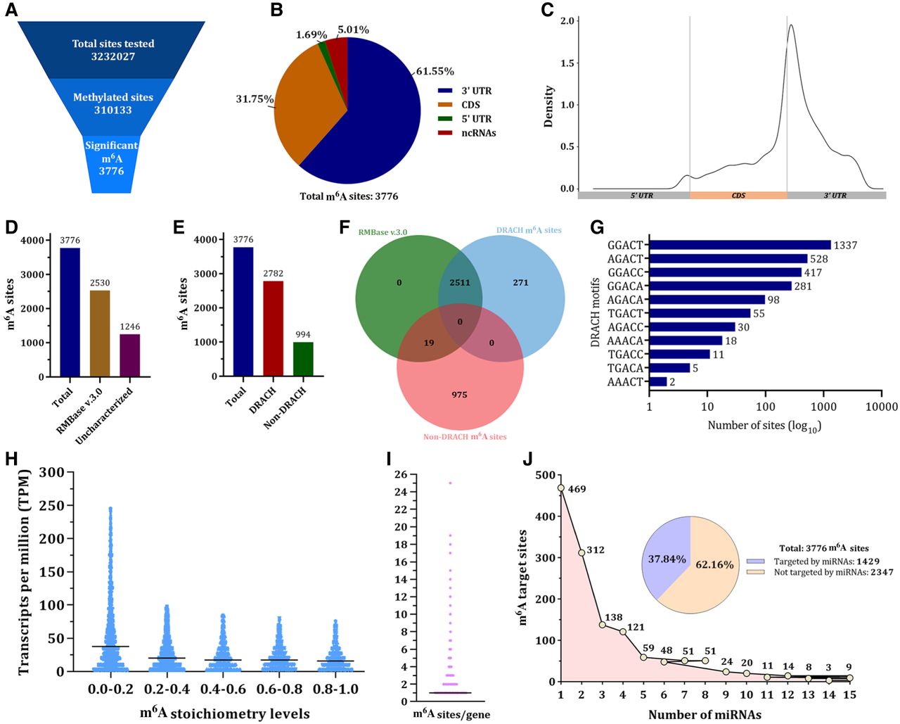
The consistency of the obtained results was validated by their direct comparison with records deposited in the RMBase v3.0, a comprehensive repository of RNA modifications detected and characterized by existing short-read high-throughput sequencing approaches, including m6A-seq and miCLIP (Xuan et al. 2024). Direct RNA sequencing data analysis highlighted 2530 m6A sites that were already deposited in RMBase v.3.0, therefore being characterized sites (Fig. 1D). Notably, the remaining 1246 detected sites represent uncharacterized m6A sites that merit further investigation. Additionally, 73.6% (2782 sites) of the identified m6A sites were found in the context of the 5′-DRACH-3′ motifs, whereas the remaining 994 sites resided in miscellaneous non-DRACH sequences (Fig. 1E). Interestingly, 90% of the high-confident 5′-DRACH-3′ sites detected in the present study are also deposited in RMBase v.3.0 and therefore represent modifications validated by antibody-based high-throughput sequencing methodologies (Fig. 1F). Motif analysis of the obtained nanopore sequencing data revealed that 5′-GGACT-3′ was the most abundant methylated 5′-DRACH-3′ motif, whereas noteworthy abundancies were also observed in the 5′-AGACT-3′, 5′-GGACC-3′, and 5′-GGACA-3′ motifs (Fig. 1G).
The final part of the m6A epitranscriptome analysis was the association between m6A methylation and transcript abundance. For this purpose, mRNA transcripts were stratified into groups based on the m6A stoichiometry levels. Our findings support that mRNA transcripts with low-stoichiometry (<0.2) m6A sites demonstrate increased expression levels compared with mRNAs with highly methylated sites (Fig. 1H). However, no significant differences were observed across other stoichiometry subgroups. Additionally, analysis showed that the m6A methylome of HeLa cells is characterized by a median value of one high-confident m6A site per gene (Fig. 1I). Finally, we investigated the overlap between high-confident m6A sites and predicted miRNA target regions. To this end, we utilized data sets from DIANA-TarBase v8 (Karagkouni et al. 2018), a curated database of experimentally validated miRNA–mRNA interactions derived from high-throughput techniques such as PAR-CLIP. Each m6A site was tested for overlap with the genomic coordinates of validated miRNA-binding sites, and the proportion of sites within miRNA target regions was quantified. Our findings support that 37.84% of the high-confident m6A sites were found to reside within experimentally supported miRNA-binding regions, highlighting the potential regulatory interplay between m6A modification and miRNA targeting (Fig. 1J).
Design of Dem6A-Vec for versatile targeted m6A demethylation
An “all-in-one” nonviral plasmid vector, Dem6A-Vec, was designed for targeted m6A demethylation (Fig. 2A). The plasmid was designed to encode the catalytically “dead” type VI-B Cas13d enzyme (mutations: R239A, H244A, R858A, and H863A) from the bacteria Ruminococcus flavefaciens (RfxCas13d) (Tang et al. 2022), fused to the characterized human m6A demethylase ALKBH5. The fused dRfxCas13d-ALKBH5 protein was tagged with both FLAG-(DYKDDDDK) and HA-(YPYDVPDYA) in the C terminus, whereas a nuclear export signal (NES) followed by a longlinker sequence was utilized for linking the two proteins and inducing the nuclear export of the fusion protein (Fig. 2A). Furthermore, the designed plasmid confers resistance to ampicillin for bacterial selection as well as resistance to puromycin for positive selection of transfected mammalian cells.
Schematic illustration of Dem6A-Vec. (A) Circular map of Dem6A-Vec. The distinct features of the plasmid are shown in different colors. The location, size, and type of each feature are also exhibited. (B) Linear map of Dem6A-Vec. The plasmid sequence encompasses two recognition sites for the type IIS PaqCI restriction endonuclease, thus enabling the scarless cloning of any RfxCas13d-compatible gRNA of choice through golden gate cloning. (C) Fluorescence microscopy images highlighting the cellular localization of Dem6A-Vec.
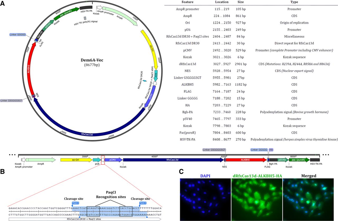
To ensure versatility in terms of targeting multiple m6A sites, Dem6A-Vec enables a U6-driven expression of a Cas13d-compatible gRNA. The gRNA cloning site of Dem6A-Vec is located downstream from the 30 nt direct repeat for RfxCas13d and includes a 32 bp sequence serving as the template region for the incorporation of any gRNA sequence through the golden gate cloning method (Ran et al. 2013). Of note, the designed plasmid contains a gRNA cloning site that encompasses two cleavage sites for the type IIS PaqCI restriction endonuclease (Kennedy et al. 2023), thus enabling the scarless cloning of the gRNA sequence into the backbone sequence of Dem6A-Vec (Fig. 2B). Finally, fluorescence microscopy highlighted that after transfection, the fused dRfxCac13d-ALKBH5 construct is expressed in both the cytoplasm and nucleus of HeLa cells (Fig. 2C).
Dem6A-Vec induces the demethylation of m6A sites in human mRNAs
To evaluate the efficiency of Dem6A-Vec in demethylating specific m6A sites in mRNAs, we implemented the designed approach on multiple candidate m6A sites that (1) were detected from direct RNA sequencing; (2) had a >0.9999 probability score from CHEUI, thus exhibiting a negligible FDR; and (3) were deposited in RMBase v.3.0 database, therefore being validated by short-read high-throughput sequencing (Supplemental Table S1). Indicatively, the present study presents detailed results of our approach on demethylating two m6A sites. The first site is located within a 5′-DRACH-3′ motif on the mRNA of the EEF2 gene (GenBank transcript ID: NM_001961.4, position: 2886), whereas the second m6A resides on a 5′-DRACH-3′ site of the RRAGA mRNA (GenBank transcript ID: NM_006570.5, position: 418). For targeted demethylation, two distinct gRNA oligonucleotide duplexes were cloned into Dem6A-Vec, and assays were performed on HeLa cells. Although EEF2 and RRAGA were selected as representative examples for initial optimization, primarily based on expression level and annotation quality, their selection was random among several high-confident m6A sites. However, to further demonstrate the versatility of the Dem6A-Vec system, multiple m6A targets with varying stoichiometries were investigated.
The demethylation efficiency of the EEF2 and RRAGA target sites was evaluated in WT cells, cells transfected with Dem6A-Vec expressing a nontargeting gRNA (NT-gRNA), and cells transfected with Dem6A-Vec expressing the gRNA specific to the m6A target. SELECT-qPCR assays were then conducted, targeting the m6A site and a nonmethylated A nucleotide of the housekeeping HPRT1 gene for normalization purposes. The principle of SELECT-qPCR, which efficiently amplifies nonmethylated A sites but exhibits significantly higher Ct values for m6A-modified sites, was employed to assess the efficiency of demethylation.
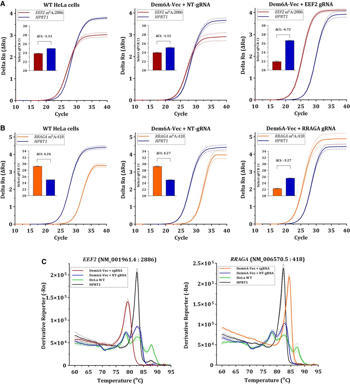
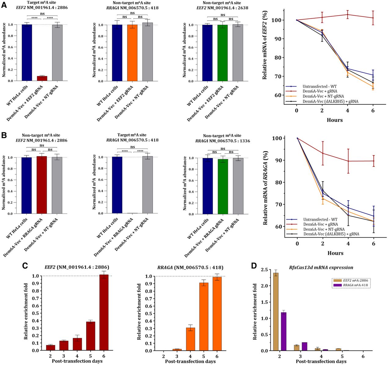
The SELECT-qPCR results showed no significant differences in normalized m6A abundance (2−ΔΔCt method) between WT cells and cells transfected with Dem6A-Vec expressing the NT-gRNA, indicating that Dem6A-Vec does not induce demethylation of the target site without the presence of site-specific gRNA. In contrast, cells transfected with Dem6A-Vec expressing the gRNA targeting the m6A:2886 site of EEF2 exhibited a notable upregulation of the produced PCR product, highlighting a successful demethylation of the target (Fig. 3A). Similar results were observed for the m6A:418 site of the RRAGA gene when the corresponding site-specific gRNA was expressed (Fig. 3B). Melt curve analysis validated the specific amplification of demethylated target sites. In contrast, WT cells and cells transfected with the NT-gRNA failed to produce PCR products, owing to the inhibitory effect of m6A on the ligation step required for successful amplification (Fig. 3C).
Demethylation impact of Dem6A-Vec at 48 h posttransfection. (A) Amplification plots from the implementation of SELECT-qPCR on the m6A:2886 of EEF2 and a nonmethylated site of the housekeeping HPRT1 gene. (B) Amplification plots from the implementation of SELECT-qPCR on the m6A:418 of RRAGA and a nonmethylated A site of the housekeeping HPRT1 gene. (C) Melt curves generated from the SELECT-qPCR assays on the EEF2 m6A:2886 and RRAGA m6A:418 sites.
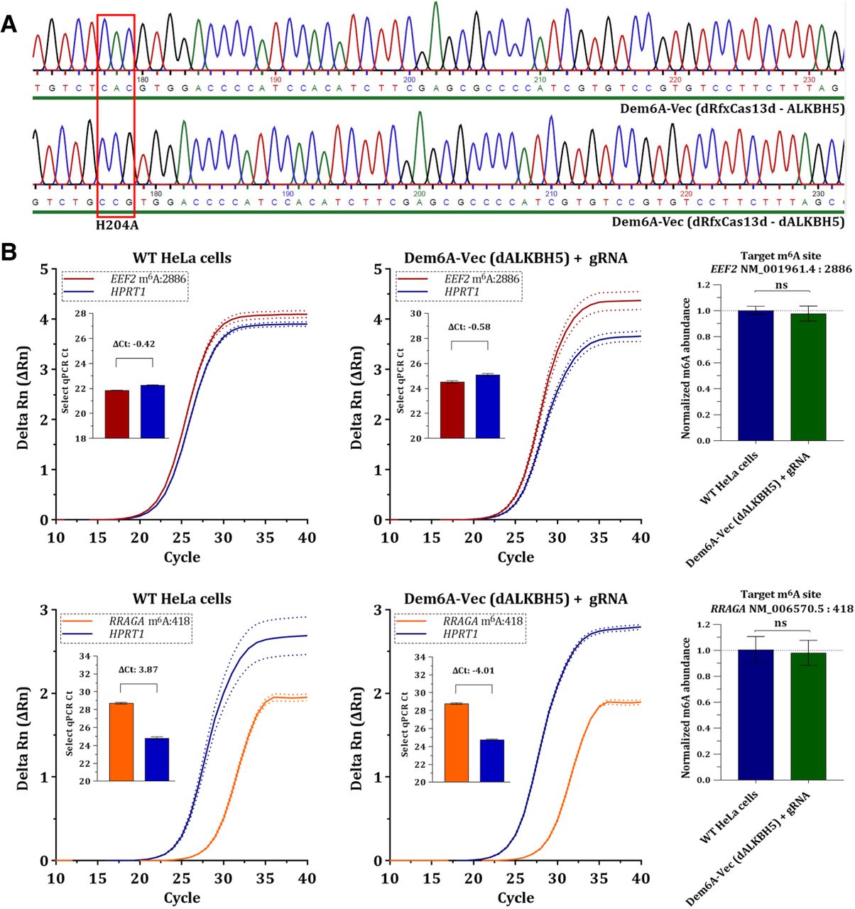
Another important aspect of the study was to rule out the possibility that m6A removal could be attributed to endogenous ALKBH5 activity rather than the expressed construct. To address this, we generated a version of Dem6A-Vec expressing a catalytically “dead” ALKBH5 variant (dALKBH5) carrying the H204A mutation (Fig. 4A). SELECT-qPCR analysis of the EEF2 and RRAGA m6A targets in HeLa cells transfected with the dALKBH5 construct revealed no significant differences compared with WT cells (Fig. 4B), confirming that demethylation depends on the enzymatic activity of the ALKBH5 in the fusion protein. Finally, to evaluate potential off-target demethylation of the designed vector across the transcriptome, we transfected cells with Dem6A-Vec expressing a NT-gRNA and performed direct RNA sequencing. Differential methylation analysis using CHEUI-diff showed no significant changes in m6A stoichiometry between the HeLa WT and transfected cells, indicating that the plasmid vector itself does not alter the global m6A methylome (Supplemental Fig. S1).
Investigation of the demethylation impact of Dem6A-Vec expressing a catalytically inactive ALKBH5 (dALKBH5) after site-directed mutagenesis leading to the H204A mutant enzyme. (A) Sanger sequencing results validating the successful generation of Dem6A-Vec expressing dALKBH5. (B) SELECT-qPCR results for m6A:2886 of EEF2 and m6A:418 of RRAGA on WT HeLa cells and cells transfected with Dem6A-Vec expressing dALKBH5. The m6A stoichiometry on both conditions was calculated based on the 2−ΔΔCt methodology, using a nonmethylated A site of HPRT1 gene as control.
To further support our findings, we performed both agarose gel electrophoresis and capillary fragment analysis of SELECT-qPCR products. Based on the capillary electrophoresis in both WT and transfected cells, the ligated amplicons that correspond to successful demethylation (∼75–80 bp) can be clearly discriminated from the nonligated products and adapter dimers (∼40 bp) for both the EEF2 and RRAGA target sites (Supplemental Fig. S2A,B). In addition, agarose electrophoresis confirms the absence of the ligated product in WT and dALKBH5-transfected cells for the EEF2 target site (Supplemental Fig. S3). These results confirm the inability of the dALKBH5-expressing construct to perform m6A editing and provide consistent findings, reinforcing the specificity of the SELECT-qPCR signal.
Dem6A-Vec induces targeted m6A demethylation without affecting adjacent m6A on the same mRNA
To investigate the specificity of Dem6A-Vec, we assessed whether demethylation was restricted to the target m6A sites (EEF2 m6A:2886 and RRAGA m6A:418) without affecting other m6A sites within the same transcript or even completely unrelated genes. For this purpose, we investigated the potential impact of the approach on the methylated EEF2 m6A:2638 site, as well as the RRAGA m6A:1336. Both these sites are within the same mRNAs as the previously demethylated target sites; they were detected by nanopore sequencing as high-confident sites and are deposited in RMBase v.3.0.
In cells transfected with the gRNA targeting EEF2 m6A:2886, 2−ΔΔCt analysis showed a significant demethylation of the target site, whereas no noteworthy changes in methylation were observed at both the EEF2 m6A:2638 and RRAGA m6A:418 sites (Fig. 5A). Similarly, in cells transfected with the gRNA targeting RRAGA m6A:418, the target site was strongly demethylated, whereas both EEF2 m6A:2886 and RRAGA m6A:1336 demonstrated nonsignificant methylation changes (Fig. 5B).
Assessment of the demethylation efficiency of Dem6A-Vec approach. (A) Targeted demethylation of the EEF2 target site (NM_001961.4 m6A:2886). Bar plots are used to demonstrate the normalized m6A abundance of the target site (EEF2 m6A:2886) and two nontarget sites, EEF2 m6A:2638 and RRAGA m6A:418. In addition, the relative expression of EEF2 mRNA is shown under actinomycin D treatment in a time line of 6 h to evaluate the mRNA stability of the target. (B) Targeted demethylation of the RRAGA target site (NM_006570.5 m6A:418). Bar plots are used to demonstrate the normalized m6A abundance of the target site (RRAGA m6A:418) and two nontarget sites, RRAGA m6A:1336 and EEF2 m6A:2886. In addition, the relative expression of RRAGA mRNA is shown under actinomycin D treatment in a time line of 6 h to evaluate the mRNA stability of the target. (C) Graphical demonstration of the normalized m6A abundance of both the EEF2 m6A:2886 and RRAGA m6A:418 sites for a 6 day posttransfection time line. (D) Normalized expression levels of the RfxCas13d mRNA expressed by Dem6A-Vec in the transfected cells for a 6 day posttransfection time line. The human housekeeping gene GAPDH was used for normalization purposes.
Demethylation of specific m6A sites by Dem6A-Vec regulates mRNA stability
Because m6A modifications on mRNAs are known to interfere with the stability of the transcribed product (Wang et al. 2014; He and He 2021; Chen et al. 2024), we investigated whether Dem6A-Vec can be utilized for assessing the impact of the target m6A on the stability of the related mRNA. For this purpose, WT and cells transfected with Dem6A-Vec expressing the NT-gRNA, as well as with the gRNA of the target, were plated and treated with actinomycin D for 6 h. Our qPCR findings demonstrated that demethylation of both EEF2 m6A:2886 and RRAGA m6A:418 leads to increased stability of the mRNA target in a time line of 6 h after actinomycin D treatment (Fig. 5A,B).
Dem6A-Vec enables reversible m6A demethylation showcasing RNA modification plasticity
In our effort to investigate the duration of the editing effects, cells were transfected, positively selected with puromycin treatment for 24 h, and collected every 24 h for SELECT-qPCR. At every time point, qPCR was also carried out to assess the mRNA levels of RfxCas13d. Interestingly, 2−ΔΔCt analysis confirmed that in both cases of the EEF2 and RRAGA target sites, the highest demethylation efficiency was achieved 48 h (2 days) posttransfection (Fig. 5C). Although both targets showed substantial demethylation on day 3, partial remethylation had already initiated. As expected, higher methylation ratios were observed for days 4 and 5, with both targets being fully remethylated on day 6. Findings on the Dem6A-Vec mRNA levels point toward the same direction, because the highest plasmid expression was observed on day 2; a notable decrease was noticed on day 3; and, on day 6, plasmid expression was decreased in a hundred-fold manner (Fig. 5D).
Dem6A-Vec impacts m6A sites with diverse methylation stoichiometries
To further evaluate the versatility of the presented approach, Dem6A-Vec was used to demethylate multiple m6A sites with varying stoichiometries and hence provide insights into the demethylation impact on low and high methylation ratios. For this purpose, gRNAs were designed and cloned to target the following sites of several random genes: CDC37 (NM_007065.4 m6A:1145), PSAT1 (NM_058179.4 m6A:1375), MRPL14 (NM_032111.4 m6A:476), LAMTOR2 (NM_014017.4 m6A:332), CDK2 (NM_001798.5 m6A:1158), and NAGLU (NM_000263.4 m6A:2002). All sites were detected by direct RNA sequencing as high-confident m6A (Supplemental Table S1). Additionally, based on the nanopore sequencing findings, CDC37 (NM_007065.4 m6A:1145) presents a minimum of 0.05 m6A stoichiometry, being the least methylated m6A in HeLa transcriptome, whereas NAGLU (NM_000263.4 m6A:2002) is a fully methylated site.
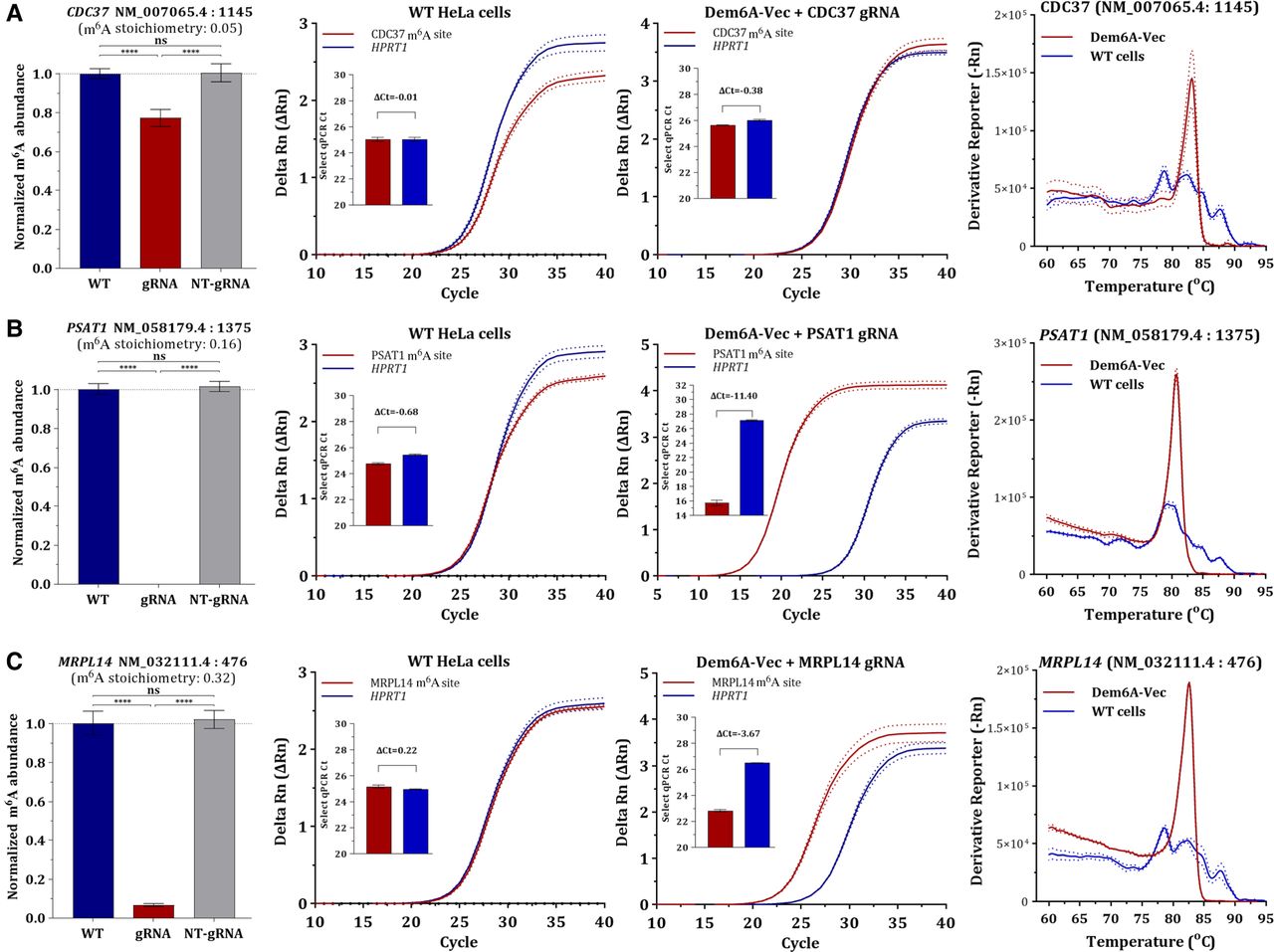
On m6A sites with low methylation ratios (stoichiometry < 0.5), Dem6A-Vec demonstrated promising results in terms of demethylation impact. In the case of the CDC37 site, which demonstrated a minimum of 0.05 stoichiometry, only a slight decrease in m6A abundancy was noticed (Fig. 6A). Despite the inherently lower baseline methylation levels, we observed noteworthy reduced m6A abundance of both the PSAT1 (m6A stoichiometry: 0.16) and MRPL14 (m6A stoichiometry: 0.32) target sites (Fig. 6B,C). Notably, our results did not identify a definitive stoichiometry threshold under which demethylation was entirely ineffective.
Evaluation of the presented targeted demethylation approach on high-confident m6A targets with <0.5 methylation stoichiometries. SELECT-qPCR data and normalized m6A abundance are shown for CDC37 (NM_007065.4 m6A:1145; A), PSAT1 (NM_058179.4 m6A:1375; B), and MRPL14 (NM_032111.4 m6A:476; C) at 48 h posttransfection with Dem6A-Vec expressing the target gRNAs.
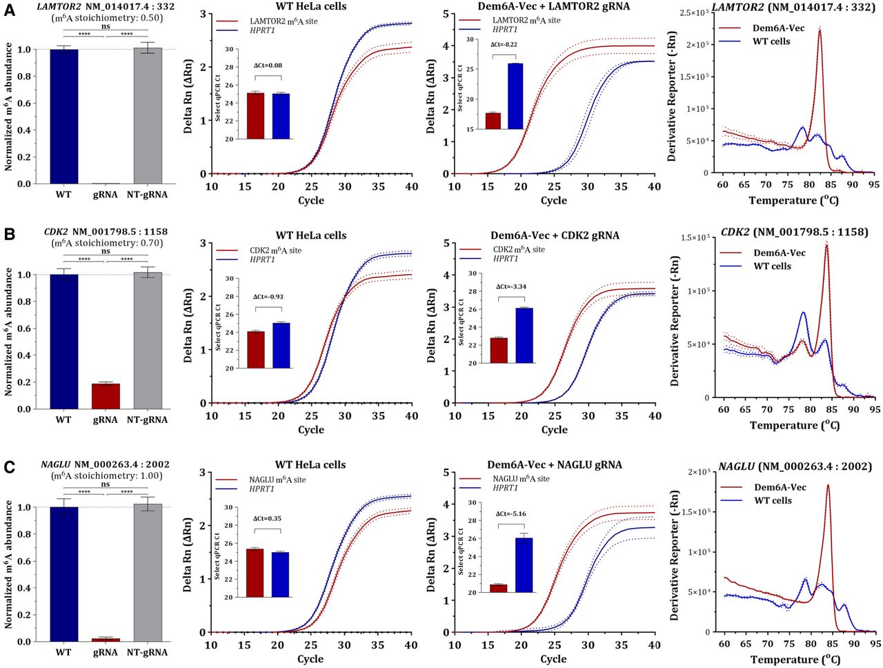
At m6A sites exhibiting high stoichiometry (>0.5), our approach induced a robust and consistent demethylation effect. Based on the 2−ΔΔCt method, the target sites of LAMTOR2 (NM_014017.4 m6A:332) and CDK2 (NM_001798.5 m6A:1158) were significantly demethylated (Fig. 7A,B). Similarly, on the fully methylated site of NAGLU (NM_000263.4 m6A:2002), Dem6A-Vec exhibited a significant reduction in methylation levels (Fig. 7C). Notably, removal of the each m6A target was also confirmed by the fact that successful generation of qPCR amplicon in the melt curve analysis was clearly observed only in the edited cells. It should be mentioned that because SYBR Green methodology was used for qPCR detection, minor signals from dimers can occasionally appear and are highly dependent on the specific primers used for the site of interest (Fig. 7B). Collectively, these findings emphasize the adaptability of Dem6A-Vec as a demethylation tool capable of targeting m6A sites irrespective of their initial stoichiometry. This versatility highlights its utility for investigating the m6A functionality in mRNAs with varying methylation dynamics.
Evaluation of the presented targeted demethylation approach on high-confident m6A targets with >0.5 methylation stoichiometries. SELECT-qPCR data and normalized m6A abundance are shown for LAMTOR2 (NM_014017.4 m6A:332; A), CDK2 (NM_001798.5 m6A:1158; B), and NAGLU (NM_000263.4 m6A:2002; C) at 48 h posttransfection with Dem6A-Vec expressing the target gRNAs.
Discussion
The field of epitranscriptomics has garnered considerable attention owing to its potential to unveil the intricate regulatory roles of RNA modifications. Among these modifications, m6A has emerged as the most abundant and dynamically regulated in eukaryotic mRNAs (Liu et al. 2020). Studies have demonstrated that m6A influences various mRNA features, including stability, splicing, export, translation, and degradation (Roundtree et al. 2017; Zaccara and Jaffrey 2020). The introduction of CRISPR/Cas13d has revolutionized epitranscriptomics by providing a robust tool for RNA processing and precise manipulation (Wessels et al. 2020). A significant advancement in this field is the development of fused constructs that integrate catalytically inactive Cas13d with effector domains, such as methyltransferases or demethylases (Konermann et al. 2018; Cao et al. 2021). This enables the specific manipulation rather than degradation of the RNA target. Although this targeted approach holds great promise for advancing research and developing novel therapeutic applications, the current number of studies utilizing tools for precise RNA manipulation is limited.
In the present study, we introduce Dem6A-Vec, an in-house developed plasmid vector designed for targeted m6A demethylation in human mRNAs. One primary advantage of Dem6A-Vec is its versatile “all-in-one” plasmid design, integrating the expression of catalytically inactive RfxCas13d fused with the major human m6A demethylase ALKBH5, as well as the U6-driven expression of any gRNA compatible with CasRx (Fig. 2A). Among the identified and studied Cas13 effector proteins, including AdmCas13d, EsCas13d, and LwaCas13a, Dem6A-Vec was designed to express RfxCas13d because this effector protein has been shown to demonstrate the most robust results in terms of targeted mRNA engineering (Konermann et al. 2018).
The design of all gRNAs utilized in the present study were based on the already identified optimal gRNA features for RfxCas13d (Xia et al. 2021). Given that RfxCas13d can, in principle, target any RNA sequence with the appropriate gRNA design, all high-confident m6A sites identified in our direct RNA sequencing data set could theoretically serve as Dem6A-Vec targets. The only technical constraint arises in cases in which a gRNA sequence contains the recognition site for the PaqCI restriction enzyme used in golden gate cloning. However, because PaqCI recognizes a 7 nt motif (5′-CACCTGC-3′), the likelihood of this occurring is extremely low. In such rare cases, alternative gRNA designs can resolve the issue, preserving the system's broad applicability. Another significant aspect of the gRNA design for the presented approach is the potential off-target activity. To assess any potential off-target activity within the target mRNA, we evaluated the methylation status of two additional m6A sites, EEF2 m6A:2638 and RRAGA m6A:1336, which are located within the same mRNAs as our target sites but were not targeted by the expressed gRNAs. SELECT-qPCR analysis revealed no significant changes in methylation at these nontarget sites. Although these results are encouraging, it is highly anticipated that any potential off-target effects would primarily depend on the sequence complementarity and specificity of the designed gRNA. Thus, transcriptome-wide off-target analysis is highly recommended for each gRNA prior to any RNA editing experiment.
In addition to HeLa cells, we sought to explore the broader applicability of Dem6A-Vec in cell types that are typically more resistant to transfection. Thus, we tested the plasmid vector in BJ and NIH/3T3 fibroblast cells and confirmed robust expression of the construct, using fluorescence microscopy. The observed results suggest that, despite the inherent challenges in transfecting primary-like and fibroblast cell lines, Dem6A-Vec can still achieve efficient delivery and expression (Supplemental Material). This expands the potential use of our tool to a wider range of biological systems and experimental contexts.
The presented approach has a differentiated design compared with previously published methods that require separate plasmids for the Cas13-ALKBH5 fusion construct and the gRNAs, necessitating multiple transfections or cotransfections and increasing the complexity of the experimental setup (Rau et al. 2019; Cao et al. 2021). The transfection of separate plasmids for the Cas-ALKBH5 fusion protein and gRNAs not only complicates the experimental workflow but also introduces variability in transfection efficiency, which may lead to inconsistent expression levels of the required components and thus may affect the efficiency and specificity of the targeted demethylation process. By incorporating these components into a single plasmid, Dem6A-Vec simplifies the process of targeted RNA editing. This “all-in-one” design ensures codelivery and coexpression of both the dRfxCas13d-ALKBH5 fusion protein and the gRNA within the same cells, thereby minimizing variability introduced by cotransfection of multiple plasmids. Although expression levels are influenced by factors such as promoter strength, the use of a single vector ensures that both components are present in the same cellular context, which is essential for the assembly and activity of the RNA-processing complex. Additionally, the unified plasmid system reduces the total amount of transfected DNA, which may help limit cytotoxicity and enhance transfection efficiency and reproducibility.
Besides enhancing the reproducibility and scalability of experiments, the use of a single plasmid vector opens new possibilities for combinatorial studies, enabling the rapid investigation of multiple m6A sites by simply altering the gRNA sequence within the plasmid. Undoubtably, this flexibility has the potential to accelerate the investigation of the functional roles of specific m6A modifications across various mRNA targets and biological contexts. To obtain candidate m6A targets for validating our approach, we implemented direct RNA sequencing on HeLa cells and performed m6A calling with CHEUI (Acera Mateos et al. 2024). However, we took into consideration only the m6A sites that demonstrated negligible FDR values (methylation probability > 0.9999) and were also deposited in the RMBase v3.0 database (Xuan et al. 2024), therefore being validated with short-read high-throughput sequencing (Supplemental Table S1).
In the present study, we demonstrated the targeted demethylation of two characterized m6A sites on the EEF2 and RRAGA mRNAs using Dem6A-Vec (Fig. 3A,B), whereas m6A targets of multiple genes (CDC37, PSAT1, MRPL14, LAMTOR2, CDK2, and NAGLU) with diverse methylation stoichiometries were used for validation of the approach. Demethylation efficiency for each investigated target was assessed based on the 2−ΔΔCt method from the SELECT-qPCR results. For demethylation assessment of EEF2 and RRAGA m6A sites in both WT and transfected HeLa cells, a targeted GLORI approach was also performed as previously described in a recent study, using reverse transcription of treated mRNA followed by nested PCR (Supplemental Fig. S4A,B; Sun et al. 2025). Although demethylation is verified by both approaches, differentiations in stoichiometry are expected mainly owing to the random nested PCR-based amplification resulting from the heavy mRNA fragmentation induced by the GLORI treatment as well as the limited quantification accuracy that SELECT-qPCR may exhibit. Specifically, for EEF2 and RRAGA m6A targets, SELECT-qPCR analysis supported that demethylation impact is fully reversible in a time line of 6 days posttransfection (Fig. 5C). Finally, our findings demonstrated that Dem6A-Vec induces targeted demethylation of m6A in mRNAs, influencing their stability, and therefore represents a robust and versatile tool for mRNA epitranscriptomics.
Another key feature of the presented approach is the choice of the restriction enzyme used for golden gate cloning of gRNAs into Dem6A-Vec. The developed approach involves the utilization of the restriction endonuclease PaqCI, which is a type IIS restriction enzyme recognized for its robustness and precision, especially in applications like golden gate assembly (Kennedy et al. 2023). PaqCI recognizes a unique 7 nt palindromic sequence, a feature that distinguishes it from many other restriction enzymes that typically recognize shorter sequences (Marillonnet and Werner 2019; Tasnim et al. 2023). Hence, the incorporation of two PaqCI cleavage sites into Dem6A-Vec for gRNA cloning is particularly beneficial in terms of modifying the plasmid vector and using different fused constructs with other effector proteins, because this longer recognition sequence reduces the chances of additional cuts in the effector protein. Consequently, although Dem6A-Vec is presented as a m6A demethylation vector, it is designed in a way that is adjustable for fusing Cas13d with other effector proteins that include methyltransferases (Wilson et al. 2020), demethylases (Li et al. 2020), or even modification-specific reader proteins (Rauch et al. 2018), enabling a wide variety of applications in epitranscriptomic research (Cox et al. 2017; Shi and Wu 2024). Specifically, the primary m6A eraser ALKBH5 that was utilized in this study can be substituted with other “writers,” “erasers,” or even “readers.” Indicatively, Dem6A-Vec could be used after the substitution of ALKBH5 with any member of the human ALKBH family because no restriction site is recognized by PaqCI. This adaptability allows researchers to expand the utility of Dem6A-Vec to investigate a wide range of RNA modifications beyond m6A, such as m5C (Zhang et al. 2024), pseudouridine, and inosine (Wang et al. 2023). Hence, by enabling the incorporation of various effectors, Dem6A-Vec provides a powerful tool for dissecting the functional roles of different RNA modifications in transcript stability, translation, and cellular homeostasis. This flexibility underscores its potential to drive discoveries across diverse fields of epitranscriptomics and RNA biology, offering opportunities to better understand the regulatory networks mediated by RNA modifications and their implications in health and disease.
The subcellular localization of Cas13d fusion proteins is increasingly recognized as a critical factor influencing RNA-processing efficiency. Although CasRx is generally cytoplasmic, its localization can shift depending on the fusion partner and the nuclear retention of crRNAs. Recent studies have demonstrated that adding an NES signal can enhance cytoplasmic activity by facilitating the export of Cas13d–crRNA complexes (Gruber et al. 2024). Based on this rationale, we incorporated a C-terminal NES into our construct to promote access to cytoplasmic mRNAs. Fluorescence microscopy confirmed that the expressed dCasRx-ALKBH5 fusion protein localizes to the cytoplasm but is also detectable in the nucleus (Fig. 2C). As various localization strategies continue to be explored, Dem6A-Vec has been designed to allow substitution of the NES with alternative signals, enabling further adaptation in future applications.
In summary, Dem6A-Vec constitutes an alternative and versatile approach for mRNA epitranscriptomic research by addressing the existing drawbacks of previous methods and providing a comprehensive and efficient approach for targeted m6A demethylation. Its integration of both the demethylase and gRNA components into a single plasmid not only simplifies the experimental process but also enhances the precision, reproducibility, and scalability of epitranscriptomic studies. By providing a versatile and efficient tool for targeted m6A demethylation, our study lays the groundwork for a deeper exploration of the functional roles of m6A modifications in gene expression and disease. The insights gained from this research have the potential to drive the development of novel therapeutic strategies targeting RNA modifications.
Methods
Cell culture
HeLa (cervical carcinoma) cells were propagated in Eagle's minimum essential medium (2 mM L-glutamine, 1 mM sodium pyruvate, 1.5 g/L NaHCO3, w: NEAA, w: EBSS), supplemented with 10% fetal bovine serum (FBS; Gibco/Life Technologies) and 1% penicillin/streptomycin (Biosera), at 37°C and 5% CO2. Dissociation of cells was performed upon 80%–90% confluency with trypsin–EDTA solution (Biowest) with a passaging ratio of 1:4.
Total RNA extraction and mRNA enrichment
The TRIzol reagent (Ambion, Thermo Fisher Scientific) was used for the total RNA isolation from HeLa cells. In the next step, mRNA enrichment was carried out with the Magnosphere UltraPure mRNA purification kit (Takara Bio), following the guidelines of the manufacturer. The quality and concentration of total and poly(A)+ RNA samples were evaluated by capillary electrophoresis on the Agilent 2100 bioanalyzer, using the RNA 6000 Pico kit (Agilent Technologies).
Direct mRNA sequencing
An initial amount of 500 ng mRNA sample was used for the library construction protocol for nanopore sequencing. For this purpose, the direct RNA sequencing kit (SQK-RNA002, Oxford Nanopore Technologies [ONT]) was used, based on the manufacturer's protocol. Briefly, the annealing and ligation of the RT adapter in the 3′ poly(A) tails of mRNAs were carried out by incubating each mRNA sample for 10 min at room temperature, using a T4 DNA ligase (New England Biolabs). Reverse transcription was performed in a 40 µL reaction volume on a Veriti 96-well fast thermal cycler (Applied Biosystems). The produced cDNA sample was purified with 1.8× Agencourt RNAClean XP beads (Beckman Coulter). The RNA adapter (RMX) was then ligated to the cDNAs in a 10 min room temperature incubation with T4 DNA ligase in a 40 µL reaction volume, and then a second purification with 0.4× Agencourt RNAClean XP beads was carried out. The final library was loaded on a FLO-MIN106D flow cell with R9.4.1 chemistry, and the sequencing run was performed on a MinION Mk1C sequencer (ONT), producing 1.22 million reads.
m6A epitranscriptome analysis
Raw sequencing data were basecalled with Guppy v.6.2.1 using the RNA model rna_r9.4.1_70bps_hac with the default read Q-score filtering. Only sequencing reads that passed the quality filtering were further processed. Basecalled sequenced reads were aligned to the reference human transcriptome (NCBI RefSeq assembly: GCF_000001405.40) using minimap2 (version 2.22) (Li 2018). Additionally, IsoQuant was employed for expression analysis and transcript quantification (Prjibelski et al. 2023). Indexing and event aligning of the basecalled reads to the reference transcriptome were conducted with nanopolish v. 0.14. Detection of m6A sites was carried out using the default pipeline parameters of CHEUI, a two-stage neural network software capable of identifying and quantifying m6A sites in nanopore RNA sequencing data sets (Acera Mateos et al. 2024). Transcriptomic sites covered with at least eight sequencing reads were taken into consideration for m6A calling. All identified m6A sites were associated with a prediction probability score, ranging from zero to one. The called m6A sites exhibiting a prediction probability > 0.9999 were considered as high-confident because they are characterized by a negligible FDR. All high-confident m6A sites detected in HeLa cells were tested on whether they were already deposited in RMBase v.3.0 (Xuan et al. 2024) and hence represented validated m6A sites with antibody based high-throughput sequencing approaches. Only high-confident m6A sites that were also deposited in RMBase v.3.0 were taken into consideration as putative m6A targets for the present methodology.
Dem6A-Vec engineering, design of gRNAs, and golden gate cloning
Engineering and delivery of Dem6A-Vec was carried out by Polyplus. Targeted demethylation of any m6A site through Dem6A-Vec requires the design of two single-stranded oligonucleotides (gRNA top and gRNA bottom). The gRNA top sequence should be complementary to the mRNA target sequence. Because the optimal gRNA design has already been elucidated for the Cas13d expressed by Dem6A-Vec (Wessels et al. 2020; Xia et al. 2021), all gRNA sequences were designed following the existing literature. In this study, gRNA top and gRNA bottom oligonucleotides were designed to target m6A sites of EEF2 and RRAGA genes, as well as several mRNA sites demonstrating differential methylation stoichiometries (Supplemental Table S2).
Annealing of the two oligos was carried out in reaction volumes of 10 µL containing 6 µL RNase-free H2O, 1 µL of each gRNA oligo (100 µM), 1 µL of 10× T4 ligation buffer (New England Biolabs), and 1 µL (10U) of T4 polynucleotide kinase (New England Biolabs). The reaction mixture was incubated for 30 min at 37°C, followed by 5 min at 95°C and a ramp down step to 25°C at 5°C/min in a Veriti 96-well fast thermal cycler (Applied Biosystems). The phosphorylated annealed oligos were diluted 1:200 in H2O, and the subsequent cloning reaction was carried out in 20 µL containing 100 ng of the designed plasmid, 2 µL of the diluted annealed oligos, 2 µL of 10× T4 ligation buffer (New England Biolabs), 1 µL (10U) of PaqCI (New England Biolabs), 0.5 µL of PaqCI activator (New England Biolabs), 1 µL of T4 DNA ligase (New England Biolabs), and H2O. The 20 µL cloning reaction was incubated for 10 cycles: 10 min at 37°C and 5 min at 16°C. In the last step, the ligation reaction was treated with exonuclease V (RecBCD) for the removal of any liner DNA fragments. For this purpose, the total ligation reaction volume of 20 µL was mixed with 5 µL 10× NEB buffer 4, 5 µL of ATP (10 mM), 1 µL (10U) of RecBCD (New England Biolabs), and 19 µL H2O to a final volume of 50 µL. Incubation was carried out for 60 min at 37°C, followed by a heat inactivation step for 30 min at 70°C. Finally, 5 µL of the RecBCD-treated reaction was mixed with 100 µL NZY5α competent cells (NZYtech) for bacterial transformation, based on the protocol of the manufacturer. The successful gRNA cloning to Dem6A-Vec was validated with Sanger sequencing using the primer 5′-GAGGGCCTATTTCCCATGATTCC-3′, which anneals to the U6 promoter sequence (Supplemental Material).
Site-directed mutagenesis
The Q5 site-directed mutagenesis kit was used to generate a Dem6A-Vec version expressing the H204A-mutated and thus catalytically inactive ALKBH5 (Zheng et al. 2013). Briefly, the first step included an exponential PCR-based amplification step using specifically designed forward (5′-CATCGTGTCTGCCGTGGACCCCATCCACATCTTC-3′) and reverse (5′-CAGCCGCCGGGCTGGTAG-3′) primers, which enabled the incorporation of the desired nucleotide substitutions. Then, the generated PCR product was subjected to kinase, ligase, and DpnI (KLD) treatment, following the protocol of the manufacturer. The treated PCR product was used for bacterial transformation, whereas validation of the successful targeted mutagenesis was performed with Sanger sequencing.
Plasmid transfection
HeLa cells were plated in six-well cell culture plates. Cells were plated 24 h prior to transfection in 3 mL complete medium per well. For each well, transfection was carried out using Opti-MEM I reduced serum medium (Gibco/Life Technologies), 4 µL Lipofectamine 3000 (Invitrogen, Thermo Fisher Scientific), 2.5 µg of plasmid DNA, and 4 µL of P3000 reagent. At 24 h after transfection, cells were treated with 2 µg/mL puromycin for the positive selection of the successfully transfected cells. The transfected cells were appropriately harvested, and total RNA extraction was performed for downstream analysis.
To evaluate the RfxCas13d mRNA expression in the transfected cells, total RNA was treated with DNase I (New England Biolabs), and then, reverse transcription was performed using random hexamers (Thermo Fisher Scientific). The generated cDNA was amplified with a qPCR-based assay using gene-specific primers for RfxCas13d (Supplemental Table S3), whereas the housekeeping gene GAPDH was used for normalization.
Fluorescence microscopy
To investigate the plasmid localization through fluorescence microscopy, an immunofluorescence application solutions kit, HA-Tag (C29F4), rabbit mAb and anti-rabbit IgG (H+L), F(ab’)2 fragment (Alexa Fluor 488 Conjugate) were purchased from Cell Signaling Technology. At 24 h postselection, HeLa cells were fixed with 4% formaldehyde for 15 min at room temperature and rinsed three times with 1× PBS for 5 min each. For immunostaining, cells were blocked using blocking buffer under shaking conditions for 60 min. Cells were then incubated with HA-tag (C29F4) rabbit mAb (1:800 dilution, Cell Signaling Technology 3724S) in antibody dilution buffer overnight at 4°C. After rinsing three times with 1× PBS, cells were incubated with fluorochrome-conjugated secondary antibody, anti-rabbit IgG (H+L), and F(ab’)2 fragment (Alexa Fluor 488 Conjugate, Cell Signaling Technology 4412S) diluted 1:600 in antibody dilution buffer for 2 h at room temperature in the dark. Finally, cells were rinsed with 1× PBS and stained with DAPI using ProLong gold antifade reagent with DAPI (Thermo Fisher Scientific P36935). Fluorescence images were captured using a Carl Zeiss Axio Vert.A1 microscope.
To evaluate the applicability of Dem6A-Vec in cell lines with lower transfection efficiency, BJ (human fibroblast) and NIH/3T3 (mouse embryonic fibroblast) cells were transfected using Lipofectamine 3000, following the same conditions as for HeLa cells. At 24 h postselection, cells were subjected to immunofluorescence staining, using anti-HA and/or anti-FLAG antibodies as described above, to confirm successful expression and localization of the Dem6A-Vec construct (Supplemental Material).
Single-base elongation and ligation-based qPCR amplification method
The already established elongation and ligation-based qPCR amplification method (SELECT-qPCR) was implemented to assess the efficiency of the presented targeted transcriptome engineering approach (Xiao et al. 2018). Based on this method, we designed two synthetic DNA oligos (UP and DOWN primers) containing adapter sequences that were designed to trap the target the m6A sites of the EEF2 and RRAGA genes (Supplemental Table S4). After ligation, site-specific amplification was performed using qPCR based on the SYBR Green method. Relative m6A abundance was calculated using the 2−ΔΔCt method, in which ΔCt was defined as the difference between the Ct value of the target site and that of the nonmethylated internal control of HPRT1, and ΔΔCt was the difference between the ΔCt of each sample and that of HeLa WT cells used as the reference condition. Relative m6A levels were then expressed as 2–ΔΔCt, enabling the evaluation of site-specific demethylation efficiency across conditions.
The initial mixtures included 1500 ng of total RNA as template, 40 nM UP primer, 40 nM DOWN primer, and 5 µM dNTPs in a total volume of 17 µL in 1× CutSmart buffer (New England Biolabs). The mixture was incubated in a hot-lid Veriti 96-well fast thermal cycler (Applied Biosystems) with the following thermal conditions: 1 min at 90°C, 1 min at 80°C, 1 min at 70°C, 1 min at 60°C, and 1 min at 50°C and then 6 min at 40°C. The final reaction mixtures were completed by adding 3 µL of 0.02 U Bst 2.0 DNA polymerase (New England Biolabs), 0.5 U SplintR ligase (New England Biolabs), and 10 nmol ATP and were incubated for 20 min at 40°C. The reaction was terminated by heat inactivation for 20 min at 80°C and stored at 4°C. For the amplification of the m6A regions of interest, qPCR assays based on the SYBR Green method were performed in a 96 fast well block QuantStudio 5 real-time PCR system (Thermo Fisher Scientific), using primers designed to anneal at the adapter sequences of UP and DOWN DNA oligos (Supplemental Table S4). Briefly, cycling conditions of qPCR reactions were as follows: 5 min at 95°C, (10 sec at 95°C; 15 sec at 60°C) repeated for 40 cycles, 15 sec at 95°C, 1 min at 60°C, 15 sec at 95°C (collecting fluorescence at a ramping rate of 0.05°C/sec), and, finally, a hold at 4°C. The qPCR data were analyzed using QuantStudio design and analysis software.
mRNA stability assay
HeLa cells were plated in 24-well plates and transfected with 0.5 µg plasmid expressing the target gRNAs and the NT-gRNA. Cells were selected with 2 µg/mL puromycin, added 24 h posttransfection. Puromycin treatment was removed after 24 h. In the next step, both transfected and WT cells were treated with actinomycin D (Gibco, Thermo Fisher Scientific) at 5 µg/mL for 0, 2, 4, and 6 h. Cells were collected and total RNA was extracted and reverse-transcribed as previously described. The first-strand cDNAs were used as templates for qPCR using gene-specific primers (Supplemental Table S3), whereas the housekeeping gene GAPDH was used for normalization.
Statistical analysis
Results are demonstrated as mean ± SD from three independent experiments. Data were analyzed by a two-tailed unpaired Student's t-test between two groups. Statistical analysis was carried out using GraphPad Prism 9. All statistical tests were two-sided (*P < 0.05, **P < 0.01, ***P < 0.001, ****P < 0.0001, ns: no significance).
Data access
The sequencing data generated in this study have been submitted to the NCBI BioProject database (https://www.ncbi.nlm.nih.gov/bioproject/) under accession number PRJNA1256807.
Competing interest statement
The authors declare no competing interests.
Acknowledgments
This research project was supported by the Hellenic Foundation for Research and Innovation (H.F.R.I.) under the “3rd Call for H.F.R.I. Research Projects to support Post-Doctoral Researchers” (project no. 07261).
Author contributions: Conceptualization, supervision, methodology, bioinformatics analysis, and writing of the original draft preparation were by P.G.A. Methodology, bioinformatics analysis, data curation, and writing of the original draft preparation were by K.A. A.S. reviewed and edited the manuscript.
Notes
[1] Supplementary material [Supplemental material is available for this article.]
[2] Article published online before print. Article, supplemental material, and publication date are at https://www.genome.org/cgi/doi/10.1101/gr.280476.125.
References
- ↵Abudayyeh OO, Gootenberg JS, Konermann S, Joung J, Slaymaker IM, Cox DB, Shmakov S, Makarova KS, Semenova E, Minakhin L, 2016. C2c2 is a single-component programmable RNA-guided RNA-targeting CRISPR effector. Science 353: aaf5573. 10.1126/science.aaf5573
- ↵Acera Mateos P, Sethi AJ, Ravindran A, Srivastava A, Woodward K, Mahmud S, Kanchi M, Guarnacci M, Xu J, Yuen WS, 2024. Prediction of m6A and m5C at single-molecule resolution reveals a transcriptome-wide co-occurrence of RNA modifications. Nat Commun 15: 3899. 10.1038/s41467-024-47953-7
- ↵Akhtar J, Lugoboni M, Junion G. 2021. m6A RNA modification in transcription regulation. Transcription 12: 266–276. 10.1080/21541264.2022.2057177
- ↵Athanasopoulou K, Boti MA, Adamopoulos PG, Skourou PC, Scorilas A. 2022. Third-generation sequencing: the spearhead towards the radical transformation of modern genomics. Life (Basel) 12: 30. 10.3390/life12010030
- ↵Azzam SK, Alsafar H, Sajini AA. 2022. FTO m6A demethylase in obesity and cancer: implications and underlying molecular mechanisms. Int J Mol Sci 23: 3800. 10.3390/ijms23073800
- ↵Bhattarai DP, Aguilo F. 2022. m6A RNA immunoprecipitation followed by high-throughput sequencing to map N6-methyladenosine. Methods Mol Biol 2404: 355–362. 10.1007/978-1-0716-1851-6_19
- ↵Cao C, Ma Q, Huang X, Li A, Liu J, Ye J, Gui Y. 2021. Targeted demethylation of the PLOD2 mRNA inhibits the proliferation and migration of renal cell carcinoma. Front Mol Biosci 8: 675683. 10.3389/fmolb.2021.675683
- ↵Chen L, Gao Y, Xu S, Yuan J, Wang M, Li T, Gong J. 2023. N6-Methyladenosine reader YTHDF family in biological processes: structures, roles, and mechanisms. Front Immunol 14: 1162607. 10.3389/fimmu.2023.1162607
- ↵Chen T, Greene GH, Motley J, Mwimba M, Luo GZ, Xu G, Karapetyan S, Xiang Y, Liu C, He C, 2024. m6A modification plays an integral role in mRNA stability and translation during pattern-triggered immunity. Proc Natl Acad Sci 121: e2411100121. 10.1073/pnas.2411100121
- ↵Cox DBT, Gootenberg JS, Abudayyeh OO, Franklin B, Kellner MJ, Joung J, Zhang F. 2017. RNA editing with CRISPR-Cas13. Science 358: 1019–1027. 10.1126/science.aaq0180
- ↵Gruber C, Krautner L, Bergant V, Grass V, Ma Z, Rheinemann L, Krus A, Reinhardt F, Mazneykova L, Rocha-Hasler M, 2024. Engineered, nucleocytoplasmic shuttling Cas13d enables highly efficient cytosolic RNA targeting. Cell Discov 10: 42. 10.1038/s41421-024-00672-1
- ↵Gupta R, Ghosh A, Chakravarti R, Singh R, Ravichandiran V, Swarnakar S, Ghosh D. 2022. Cas13d: a new molecular scissor for transcriptome engineering. Front Cell Dev Biol 10: 866800. 10.3389/fcell.2022.866800
- ↵He PC, He C. 2021. m6A RNA methylation: from mechanisms to therapeutic potential. EMBO J 40: e105977. 10.15252/embj.2020105977
- ↵Karagkouni D, Paraskevopoulou MD, Chatzopoulos S, Vlachos IS, Tastsoglou S, Kanellos I, Papadimitriou D, Kavakiotis I, Maniou S, Skoufos G, 2018. DIANA-TarBase v8: a decade-long collection of experimentally supported miRNA-gene interactions. Nucleic Acids Res 46: D239–D245. 10.1093/nar/gkx1141
- ↵Kennedy MA, Hosford CJ, Azumaya CM, Luyten YA, Chen M, Morgan RD, Stoddard BL. 2023. Structures, activity and mechanism of the type IIS restriction endonuclease PaqCI. Nucleic Acids Res 51: 4467–4487. 10.1093/nar/gkad228
- ↵Konermann S, Lotfy P, Brideau NJ, Oki J, Shokhirev MN, Hsu PD. 2018. Transcriptome engineering with RNA-targeting type VI-D CRISPR effectors. Cell 173: 665–676.e14. 10.1016/j.cell.2018.02.033
- ↵Kretschmer J, Rao H, Hackert P, Sloan KE, Höbartner C, Bohnsack MT. 2018. The m6A reader protein YTHDC2 interacts with the small ribosomal subunit and the 5′-3′ exoribonuclease XRN1. RNA 24: 1339–1350. 10.1261/rna.064238.117
- ↵Leger A, Amaral PP, Pandolfini L, Capitanchik C, Capraro F, Miano V, Migliori V, Toolan-Kerr P, Sideri T, Enright AJ, 2021. RNA modifications detection by comparative nanopore direct RNA sequencing. Nat Commun 12: 7198. 10.1038/s41467-021-27393-3
- ↵Li H. 2018. Minimap2: pairwise alignment for nucleotide sequences. Bioinformatics 34: 3094–3100. 10.1093/bioinformatics/bty191
- ↵Li HB, Tong J, Zhu S, Batista PJ, Duffy EE, Zhao J, Bailis W, Cao G, Kroehling L, Chen Y, 2017. m6A mRNA methylation controls T cell homeostasis by targeting the IL-7/STAT5/SOCS pathways. Nature 548: 338–342. 10.1038/nature23450
- ↵Li J, Chen Z, Chen F, Xie G, Ling Y, Peng Y, Lin Y, Luo N, Chiang CM, Wang H. 2020. Targeted mRNA demethylation using an engineered dCas13b-ALKBH5 fusion protein. Nucleic Acids Res 48: 5684–5694. 10.1093/nar/gkaa269
- ↵Liu J, Dou X, Chen C, Chen C, Liu C, Xu MM, Zhao S, Shen B, Gao Y, Han D, 2020. N6-methyladenosine of chromosome-associated regulatory RNA regulates chromatin state and transcription. Science 367: 580–586. 10.1126/science.aay6018
- ↵Liu H, Zheng J, Liao A. 2022. The regulation and potential roles of m6A modifications in early embryonic development and immune tolerance at the maternal-fetal interface. Front Immunol 13: 988130. 10.3389/fimmu.2022.988130
- ↵Marillonnet S, Werner S. 2019. Assembly of complex pathways using type IIs restriction enzymes. Methods Mol Biol 1927: 93–109. 10.1007/978-1-4939-9142-6_7
- ↵McIntyre ABR, Gokhale NS, Cerchietti L, Jaffrey SR, Horner SM, Mason CE. 2020. Limits in the detection of m6A changes using MeRIP/m6A-seq. Sci Rep 10: 6590. 10.1038/s41598-020-63355-3
- ↵Meyer KD, Jaffrey SR. 2014. The dynamic epitranscriptome: N6-methyladenosine and gene expression control. Nat Rev Mol Cell Biol 15: 313–326. 10.1038/nrm3785
- ↵Molinie B, Giallourakis CC. 2017. Genome-wide location analyses of N6-methyladenosine modifications (m6A-seq). Methods Mol Biol 1562: 45–53. 10.1007/978-1-4939-6807-7_4
- ↵Nachtergaele S, He C. 2017. The emerging biology of RNA post-transcriptional modifications. RNA Biol 14: 156–163. 10.1080/15476286.2016.1267096
- ↵Nombela P, Miguel-López B, Blanco S. 2021. The role of m6A, m5C and Psi RNA modifications in cancer: novel therapeutic opportunities. Mol Cancer 20: 18. 10.1186/s12943-020-01263-w
- ↵Pickar-Oliver A, Gersbach CA. 2019. The next generation of CRISPR-Cas technologies and applications. Nat Rev Mol Cell Biol 20: 490–507. 10.1038/s41580-019-0131-5
- ↵Prjibelski AD, Mikheenko A, Joglekar A, Smetanin A, Jarroux J, Lapidus AL, Tilgner HU. 2023. Accurate isoform discovery with IsoQuant using long reads. Nat Biotechnol 41: 915–918. 10.1038/s41587-022-01565-y
- ↵Ramesh-Kumar D, Guil S. 2022. The IGF2BP family of RNA binding proteins links epitranscriptomics to cancer. Semin Cancer Biol 86: 18–31. 10.1016/j.semcancer.2022.05.009
- ↵Ran FA, Hsu PD, Wright J, Agarwala V, Scott DA, Zhang F. 2013. Genome engineering using the CRISPR-Cas9 system. Nat Protoc 8: 2281–2308. 10.1038/nprot.2013.143
- ↵Rau K, Rösner L, Rentmeister A. 2019. Sequence-specific m6A demethylation in RNA by FTO fused to RCas9. RNA 25: 1311–1323. 10.1261/rna.070706.119
- ↵Rauch S, He C, Dickinson BC. 2018. Targeted m6A reader proteins to study epitranscriptomic regulation of single RNAs. J Am Chem Soc 140: 11974–11981. 10.1021/jacs.8b05012
- ↵Roundtree IA, Evans ME, Pan T, He C. 2017. Dynamic RNA modifications in gene expression regulation. Cell 169: 1187–1200. 10.1016/j.cell.2017.05.045
- ↵Shan T, Liu F, Wen M, Chen Z, Li S, Wang Y, Cheng H, Zhou Y. 2023. m6A modification negatively regulates translation by switching mRNA from polysome to P-body via IGF2BP3. Mol Cell 83: 4494–4508.e6 10.1016/j.molcel.2023.10.040
- ↵Shi P, Wu X. 2024. Programmable RNA targeting with CRISPR-Cas13. RNA Biol 21: 575–583. 10.1080/15476286.2024.2351657
- ↵Stephenson W, Razaghi R, Busan S, Weeks KM, Timp W, Smibert P. 2022. Direct detection of RNA modifications and structure using single-molecule nanopore sequencing. Cell Genom 2: 100097. 10.1016/j.xgen.2022.100097
- ↵Sun H, Lu B, Zhang Z, Xiao Y, Zhou Z, Xi L, Li Z, Jiang Z, Zhang J, Wang M, 2025. Mild and ultrafast GLORI enables absolute quantification of m6A methylome from low-input samples. Nat Methods 22: 1226–1236. 10.1038/s41592-025-02680-9
- ↵Tan X, Zheng C, Zhuang Y, Jin P, Wang F. 2023. The m6A reader PRRC2A is essential for meiosis I completion during spermatogenesis. Nat Commun 14: 1636. 10.1038/s41467-023-37252-y
- ↵Tang XE, Tan SX, Hoon S, Yeo GW. 2022. Pre-existing adaptive immunity to the RNA-editing enzyme Cas13d in humans. Nat Med 28: 1372–1376. 10.1038/s41591-022-01848-6
- ↵Tasnim M, Selph TJ, Olcott J, Hill JT. 2023. The type IIS restriction enzyme MmeI can cut across a double-strand break. Mol Biol Rep 50: 5495–5499. 10.1007/s11033-023-08375-8
- ↵Terns MP. 2018. CRISPR-based technologies: impact of RNA-targeting systems. Mol Cell 72: 404–412. 10.1016/j.molcel.2018.09.018
- ↵Wang X, Lu Z, Gomez A, Hon GC, Yue Y, Han D, Fu Y, Parisien M, Dai Q, Jia G, 2014. N6-Methyladenosine-dependent regulation of messenger RNA stability. Nature 505: 117–120. 10.1038/nature12730
- ↵Wang J, Wang J, Gu Q, Ma Y, Yang Y, Zhu J, Zhang Q. 2020. The biological function of m6A demethylase ALKBH5 and its role in human disease. Cancer Cell Int 20: 347. 10.1186/s12935-020-01450-1
- ↵Wang X, Zhang R, Yang D, Li G, Fan Z, Du H, Wang Z, Liu Y, Lin J, Wu X, 2023. Develop a compact RNA base editor by fusing ADAR with engineered EcCas6e. Adv Sci (Weinh) 10: e2206813. 10.1002/advs.202206813
- ↵Wessels HH, Méndez-Mancilla A, Guo X, Legut M, Daniloski Z, Sanjana NE. 2020. Massively parallel Cas13 screens reveal principles for guide RNA design. Nat Biotechnol 38: 722–727. 10.1038/s41587-020-0456-9
- ↵Wilson C, Chen PJ, Miao Z, Liu DR. 2020. Programmable m6A modification of cellular RNAs with a Cas13-directed methyltransferase. Nat Biotechnol 38: 1431–1440. 10.1038/s41587-020-0572-6
- ↵Xia Z, Tang M, Ma J, Zhang H, Gimple RC, Prager BC, Tang H, Sun C, Liu F, Lin P, 2021. Epitranscriptomic editing of the RNA N6-methyladenosine modification by dCasRx conjugated methyltransferase and demethylase. Nucleic Acids Res 49: 7361–7374. 10.1093/nar/gkab517
- ↵Xiang Y, Laurent B, Hsu CH, Nachtergaele S, Lu Z, Sheng W, Xu C, Chen H, Ouyang J, Wang S, 2017. RNA m6a methylation regulates the ultraviolet-induced DNA damage response. Nature 543: 573–576. 10.1038/nature21671
- ↵Xiao Y, Wang Y, Tang Q, Wei L, Zhang X, Jia G. 2018. An elongation- and ligation-based qPCR amplification method for the radiolabeling-free detection of locus-specific N6-methyladenosine modification. Angew Chem Int Ed Engl 57: 15995–16000. 10.1002/anie.201807942
- ↵Xiong X, Yi C, Peng J. 2017. Epitranscriptomics: toward a better understanding of RNA modifications. Genomics Proteomics Bioinformatics 15: 147–153. 10.1016/j.gpb.2017.03.003
- ↵Xu Y, Zhang Y, Luo Y, Qiu G, Lu J, He M, Wang Y. 2023. Novel insights into the METTL3-METTL14 complex in musculoskeletal diseases. Cell Death Discov 9: 170. 10.1038/s41420-023-01435-9
- ↵Xuan J, Chen L, Chen Z, Pang J, Huang J, Lin J, Zheng L, Li B, Qu L, Yang J. 2024. RMBase v3.0: decode the landscape, mechanisms and functions of RNA modifications. Nucleic Acids Res 52: D273–D284. 10.1093/nar/gkad1070
- ↵Yang Z, Zhang S, Xiong J, Xia T, Zhu R, Miao M, Li K, Chen W, Zhang L, You Y, 2024. The m6A demethylases FTO and ALKBH5 aggravate the malignant progression of nasopharyngeal carcinoma by coregulating ARHGAP35. Cell Death Discov 10: 43. 10.1038/s41420-024-01810-0
- ↵Zaccara S, Jaffrey SR. 2020. A unified model for the function of YTHDF proteins in regulating m6A-modified mRNA. Cell 181: 1582–1595.e18. 10.1016/j.cell.2020.05.012
- ↵Zhang Y, Lu L, Li X. 2022. Detection technologies for RNA modifications. Exp Mol Med 54: 1601–1616. 10.1038/s12276-022-00821-0
- ↵Zhang T, Zhao F, Li J, Sun X, Zhang X, Wang H, Fan P, Lai L, Li Z, Sui T. 2024. Programmable RNA 5-methylcytosine (m5C) modification of cellular RNAs by dCasRx conjugated methyltransferase and demethylase. Nucleic Acids Res 52: 2776–2791. 10.1093/nar/gkae110
- ↵Zheng G, Dahl JA, Niu Y, Fedorcsak P, Huang CM, Li CJ, Vågbø CB, Shi Y, Wang WL, Song SH, 2013. ALKBH5 is a mammalian RNA demethylase that impacts RNA metabolism and mouse fertility. Mol Cell 49: 18–29. 10.1016/j.molcel.2012.10.015
- ↵Zheng P, Zhou C, Ding Y, Liu B, Lu L, Zhu F, Duan S. 2023. Nanopore sequencing technology and its applications. MedComm (2020) 4: e316. 10.1002/mco2.316
- ↵Zhou J, Wan J, Gao X, Zhang X, Jaffrey SR, Qian SB. 2015. Dynamic m6A mRNA methylation directs translational control of heat shock response. Nature 526: 591–594. 10.1038/nature15377
- ↵Zhou Z, Lv J, Yu H, Han J, Yang X, Feng D, Wu Q, Yuan B, Lu Q, Yang H. 2020. Mechanism of RNA modification N6-methyladenosine in human cancer. Mol Cancer 19: 104. 10.1186/s12943-020-01216-3
- ↵Zhu ZM, Huo FC, Zhang J, Shan HJ, Pei DS. 2023. Crosstalk between m6A modification and alternative splicing during cancer progression. Clin Transl Med 13: e1460. 10.1002/ctm2.1460