

Abstract
Somatic hypermutation (SHM) drives antibody affinity maturation in B cells. By mimicking this process, guided hypermutation (GHM) tools employing CRISPR systems and activation-induced cytidine deaminase (AID) have advanced antibody development. However, GHM-induced mutations in cultured cells exhibit mutation patterns distinct from those observed in natural antibody diversification following in vivo affinity selection. To address this, we engineer a hyper-antibody editor, HAE1, by integrating cytidine and adenine deaminases with a nicked, PAMless Cas9 variant, SpRY, to closely resemble the mutation spectrum of natural SHM. Moreover, to streamline mutation, selection, and validation within the same cells, we develop a dual-expression system in HEK293F cells that allows simultaneous expression of both transmembrane and secreted full-length antibodies. Using this system, we apply HAE1 to the SARS-CoV-2 neutralizing antibody CV07-209 and restore the antibody's binding affinity and neutralization potency against Omicron variants, specifically BA.1, including at least one mutation beyond the reach of current GHM tools. HAE1 thus provides a versatile, high-throughput strategy for expediting antibody evolution, presenting significant potential for therapeutic antibody development and protein engineering.
The development of antibody therapies has revolutionized the treatment of various diseases, including viral infections, cancer, and autoimmune disorders (Mullard 2021; Carter and Rajpal 2022; Paul et al. 2024). The efficacy of these treatments is highly dependent on the affinity of antibodies against therapeutic targets (Oostindie et al. 2022). However, a significant challenge in antibody therapy is immune escape due to rapid antigen mutations (Hegde and Chen 2020; Cao et al. 2022; Lin et al. 2024). To address this issue, rapid evolution is a crucial strategy for the development of effective therapeutic antibodies against mutated antigens (Desautels et al. 2024; Peled et al. 2024; Bai et al. 2025). Naturally, affinity maturation is achieved through somatic hypermutation (SHM) in B cells following antigen stimulation in germinal centers (GCs) (Victora and Nussenzweig 2012; Alt et al. 2013). During SHM, the activation-induced cytidine deaminase (AID) introduces point mutations within the variable regions of antibody heavy and light chains, mainly in the antigen-binding complementarity determining regions (CDRs) (Di Noia and Neuberger 2007; Peled et al. 2008; Yeap et al. 2015; Wang et al. 2023).
Incorporating antibody genes into the immunoglobulin (Ig) loci of mouse B cells allows endogenous AID to drive affinity maturation of human antibodies effectively upon antigen immunization (Nie et al. 2020; Nahmad et al. 2022; He et al. 2023; Yin et al. 2024), but these approaches require the development of specialized mouse models. Alternatively, classical directed evolution and continuous evolution methods have been applied to optimize antibody affinity in vitro or in cultured cells (Molina et al. 2022). Classical directed evolution relies on random mutagenesis techniques including error-prone PCR and saturated mutations to construct and display antibody libraries in phage or yeast (Hawkins et al. 1992; Chao et al. 2006). In contrast, continuous evolution requires a hypermutation system that induces mutagenesis autonomously in growing cells (Hendel and Shoulders 2021; Molina et al. 2022). AID is broadly involved in these systems to mimic SHM, either through endogenous expression in B cell lines (e.g., DT40, Ramos) (Cumbers et al. 2002; Voss et al. 2019; Masuda et al. 2022) or through ectopic expression in non-B cells (Martin and Scharff 2002; Bowers et al. 2011; Chen et al. 2012; Luo et al. 2020). However, the mutation rates and diversity achieved through these methods are quite limited in comparison to those in the GC B cells (Martin et al. 2002; Martin and Scharff 2002; Luo et al. 2020).
Leveraging the deaminase activity of AID or similar enzymes, base editors have been developed to efficiently induce mutations in target genes (Hess et al. 2017; Rees and Liu 2018). Specifically, deoxynucleotide deaminase was recruited to the target site by a nicked variant of SpCas9 (nCas9) and single guide RNAs (sgRNAs) to promote nucleotide substitutions. Initially, members of the APOBEC deaminase family, such as AID, APOBEC1, and APOBEC3A, were used to create cytosine base editors (CBEs) to induce C-to-T mutations (Komor et al. 2016). Later, the engineered TadA enzyme was introduced to develop the adenine base editors (ABEs), enabling A-to-G mutations (Gaudelli et al. 2017; Richter et al. 2020). Dual base editors, combining both types of deaminases, allowed simultaneous A-to-G and C-to-T mutations (Grünewald et al. 2020; Wu et al. 2020; Zhang et al. 2020; Liang et al. 2022). Inspired by the efficient targeting and mutagenesis of base editors, guided hypermutation (GHM) tools combining hyperactive AID mutants and nuclease-dead Cas9 (dCas9) have been used to target antibody variable regions to enhance the antibody affinity in vitro. The GHM tools enhance the mutation efficiency of conventional antibody evolution but inherit the key limitation of restricted mutation spectrum compared to natural SHM (Hess et al. 2016; Liu et al. 2018; Devilder et al. 2019).
In this study, we develop a robust GHM tool, termed hyper-antibody editor 1 (HAE1), capable of targeting any given CDR sequence to generate mutation patterns that resemble those of natural SHM. Using HAE1 and a designed mammalian dual-expression system, we successfully extended the neutralizing potency of CV07-209 against viral escape variants.
Results
Development of HAE1 for broad mutation spectrum
The NGG-protospacer adjacent motif (PAM) of conventional SpCas9 limits the design of target sites on antibody CDRs, which has been partially compensated by the MS2 system used in previous tools such as DBE and CRISPR-X (Hess et al. 2016; Liu et al. 2018; Devilder et al. 2019). However, the near-NNN PAM recognition of SpRY enables almost complete coverage of any given target sequence (Walton et al. 2020). For instance, about 47 programmable sgRNAs could be designed to target the CDR1 and CDR2 of each human IGHV gene (Manso et al. 2022) by SpRY, more than nine times of SpCas9 and two times of another PAM-flexible SpCas9 mutant, SpG (NG PAM) (Supplemental Fig. S1A). Therefore, we fused a highly active AID variant (AIDmono) (Qiao et al. 2017) with a nickase variant SpRY-D10A, developing the PAMless-AID. Eighteen sites were designed to target the integrated eGFP gene in HEK293T cells, including five NAN, four NGN, four NCN, and five NTN PAMs (Fig. 1A). PAMless-AID achieved an average substitution efficiency of ∼35.0% (3.6% to 78.0%) with NRN (R = A or G) PAM showing a higher substitution efficiency compared to NYN (Y = C or T) PAM (Fig. 1B), consistent with the previous study (Walton et al. 2020). Moreover, the mutation spectrum of PAMless-AID is predominantly concentrated in C:G base pairs, with 92% C:G mutations and 8% A:T mutations (Fig. 1C), similar to DBE and CRISPR-X (Hess et al. 2016; Liu et al. 2018; Devilder et al. 2019). In contrast, human antibody repertoires exhibit a border mutation spectrum, with 57% C:G mutations and 43% A:T mutations (Fig. 1D; Di Noia and Neuberger 2007; Briney et al. 2019). In this regard, the PAMless-AID and similar GHM systems require further optimization to better mimic the natural antibody diversity.
Development of the hyper-antibody editor. (A) Schematics for the assessment of editing efficiency for PAMless-AID and HAE1. 293T-eGFP cells were cotransfected with various sgRNAs and either PAMless-AID or HAE1. BFP-positive cells were sorted 72 h posttransfection for genomic DNA extraction and amplicon sequencing. Editing efficiency and mutation spectrums were analyzed by CRISPResso2. (B) Substitution frequencies of PAMless-AID at 18 sites with NNN-PAM targeting eGFP. Numbers above each bar represent the median substitution percentage. Whiskers extend from the minimum to the maximum values, and box borders represent the first and third quartiles. For NAN and NTN, n = 5; for NCN and NGN, n = 4. (C) The mutation spectrum generated by PAMless-AID at 18 sites targeting eGFP. The entire sequence covered by sgRNAs (20 bp) is calculated for each type of base substitution (each type of substitution divided by total substitutions of four nucleotides occurring within the 20-bp region). The heat map shows the average value of each substitution type of 18 sites. (D) The mutation spectrum generated by AID in human B cells. (E) Substitution efficiencies of HAE1 with different deaminase mutations across 18 sites targeting eGFP. Statistical analysis was performed using a paired two-tailed t-test, with P values indicated or noted as not significant (n.s.) if P > 0.05. (F) The mutation spectrum generated by HAE1 at 18 sites targeting eGFP. (G) Editing window of HAE1. The y-axis indicates the substitution frequencies of C and A at each nucleotide across 18 sites targeting eGFP (n ≥ 3 at each nucleotide). Data are presented as mean ± SEM.
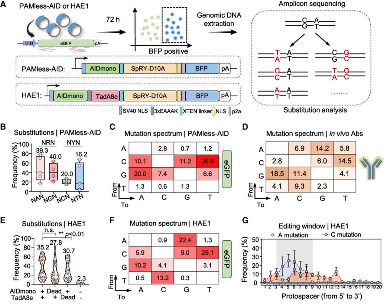
To broaden the mutation spectrum, we incorporated the A-to-G mutator TadA8e into the PAMless-AID system, developing the hyper-antibody editor 1 (Fig. 1A). TadA8e is one of the most highly active adenine deaminase, capable of functioning as a monomer (Richter et al. 2020). Although TadA8e performance remained comparable, directly fused AIDmono in HAE1 showed significantly higher activity compared with the MS2-recruited AID (Supplemental Fig. S1B,C). HAE1 generates efficient substitutions with 61% C:G mutations and 39% A:T mutations, similar to the natural antibody repertoire (Fig. 1E,F). Inactivating either AIDmono or TadA8e within HAE1 induces robust substitutions with a biased mutation spectrum, indicating that AIDmono and TadA8e function cooperatively (Supplemental Fig. S1D,E). Moreover, HAE1 induces ∼11.0% deletions and 2.2% insertions, both dependent on AIDmono (Supplemental Fig. S1F), resembling the features of natural antibody repertoires (Hao et al. 2023). HAE1 demonstrated efficient A and C mutagenesis within a 7-nt editing window (positions 3–9), combining A mutations (positions 3–9) induced by TadA8e and a broader C mutation (positions 2–13) induced by AIDmono (Fig. 1G; Supplemental Fig. S1G). These results imply that HAE1 is an efficient GHM tool with an enlarged mutation range for antibody evolution.
Development of an antibody dual-expression platform in HEK293F cells
Neutralizing antibodies targeting the spike (S) protein of SARS-CoV-2 have proven to be an effective therapeutic strategy throughout the COVID-19 pandemic (Taylor et al. 2021). Given the rapid antigenic evolution of the virus, adapting existing antibodies to recognize emerging variants provides an efficient route for neutralizing antibody development compared to discovering new candidates from convalescent individuals (Kuramochi et al. 2022; Desautels et al. 2024; Hie et al. 2024). Through analyzing the coronavirus antibody database (CoV-AbDab) (Raybould et al. 2021), we identified 447 loss-of-function human antibodies against Omicron variants. Because germline-like antibodies with low SHM have greater potential for affinity maturation against novel variants (Supplemental Fig. S2A; Muecksch et al. 2021), we focused on 13 antibodies with low SHM frequency (≤1 amino acid [aa]) and without structural information. CV07-209 emerged as the most promising candidate for further evolution due to its high affinity against wild-type SARS-CoV-2 and the absence of self-reactivity (Fig. 2A). CV07-209 belongs to the group B IgG-neutralizing antibodies and was originally isolated from an early patient infected with SARS-CoV-2 (Kreye et al. 2020; Cao et al. 2022).
Establishment of the dual-expression system. (A) A schematic diagram outlining the screening conditions of antibodies (Abs) from Cov-AbDab for antibody evolution. (B) A schematic diagram showing the dual-expression system mediated by alternative splicing. The variable (V) and constant regions (C) of the heavy chain are depicted as VH and CH. The expression of transmembrane or secreted antibodies is regulated by constructs in their spliced or unspliced forms, respectively. Asterisk (*), stop codon; TM, transmembrane domain. (C) Relative splicing efficiency of the indicated introns. Expression level is normalized to transmembrane (tmAb) and secreted (sAb) antibodies, respectively. Data are presented as mean ± SEM from three biological replicates. (D) Left: FACS data showing the expression of transmembrane antibodies and their binding to Delta (top) and BA.1 S protein (bottom). Right: ELISA results with supernatant indicating the expression levels of secreted antibodies and their response signals to Delta (top) and BA.1 S protein (bottom).
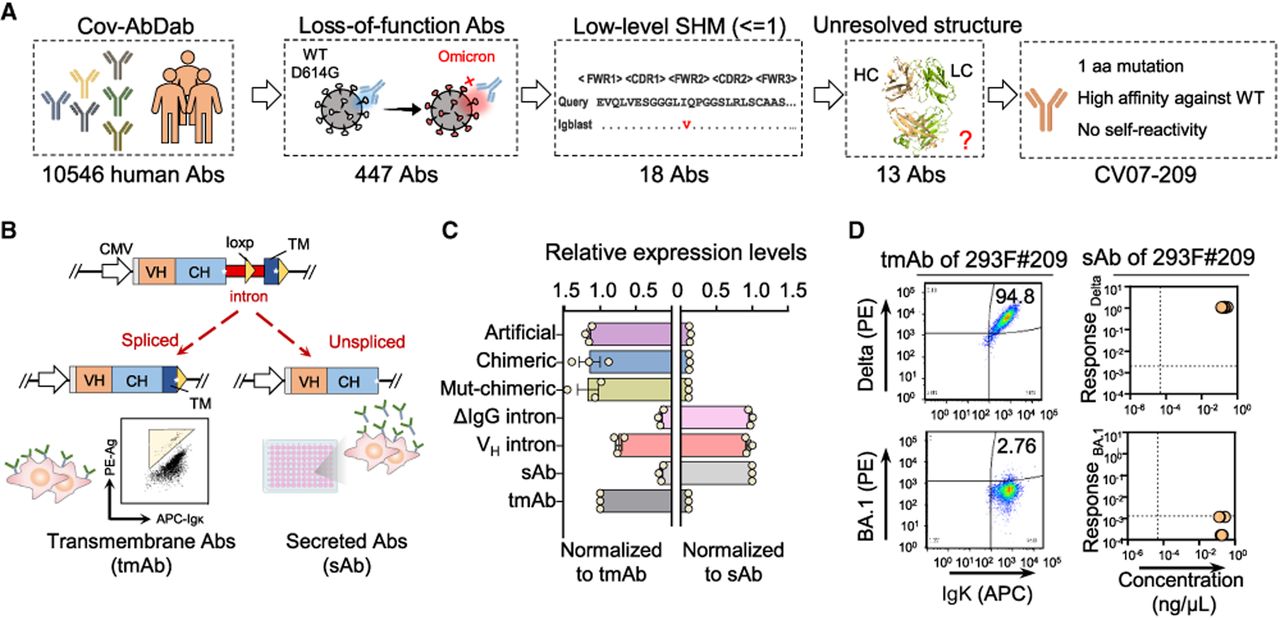
To streamline antibody evolution and validation in mammalian cells, we sought to develop a dual-expression system that supports expression of both transmembrane and secreted antibodies. The transmembrane form allows efficient cell isolation via fluorescence-activated cell sorting (FACS), whereas the secreted form facilitates antibody validation through enzyme-linked immunosorbent assay (ELISA) using the supernatants. For this purpose, we inserted a universal transmembrane motif from PDGFRB at the 3′ end of the antibody gene, flanked by two codirectional LoxP sites, enabling cleavage by Cre recombinase (Fig. 2B). We tested five different introns to accommodate the upstream LoxP site, including an artificial intron (Guzzardo et al. 2017), a chimeric intron from CAG promotor, a mutated form of the chimeric intron (mut-chimeric), a truncated natural BCR pre-mRNA intron between CH3 and IgG-TM (IgG intron), and a short intron between the VH leader exons (VH intron). We found the VH intron supported simultaneous efficient expression of both transmembrane and secreted forms of the CV07-209 antibody before Cre treatment (Fig. 2C). Therefore, we integrated the CV07-209 gene with the VH intron and PDGFRB transmembrane motif into the AAVS1 locus of HEK293F cells (Supplemental Fig. S2B). The resulting stable cell line (293F#209) was efficiently labeled by the Delta but not the BA.1 S protein. Similarly, culture supernatants containing secreted antibodies could recognize the Delta but not the BA.1 S protein (Fig. 2D), indicating the potential of 293F#209 cells for both antibody evolution and validation.
Profiling antibody evolution efficiency by high-throughput sequencing
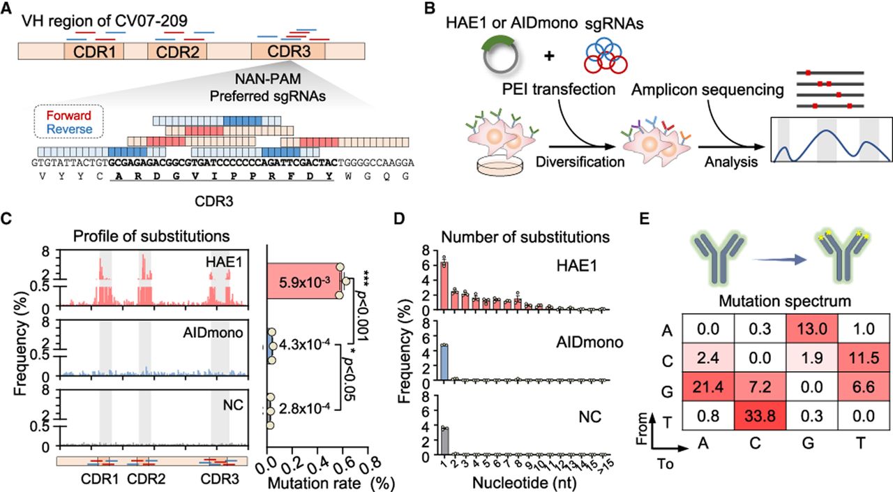
To assess the efficiency of HAE1 in antibody evolution, we targeted the heavy chain variable region (VH region) of the CV07-209 gene in 293F#209 cells (Supplemental Fig. S3A). We designed 14 sgRNAs with NNN-PAM (12 NRN and two NYN), including four targeting CDR1, four targeting CDR2, and six targeting CDR3. Each sgRNA was optimized to place the CDR residues within the ideal editing window (positions 3–9 bp) (Figs. 1G, 3A). Transient HAE1 expression was induced by cotransfection of the HAE1 and a mixture of sgRNA plasmids into 293F#209 cells using polyethyleneimine. The AIDmono was overexpressed as a control. Cells were collected 72 h posttransfection without enrichment, and the genomic DNA was extracted for mutation profiling via amplicon sequencing (Fig. 3B). HAE1 achieved a total substitution frequency of 19.9%, whereas AIDmono displayed very low activity (5.0%), only slightly above the background levels (3.7%) (Supplemental Fig. S3B). The substitutions induced by HAE1 were predominantly concentrated within the CDRs targeted by sgRNAs, with CDR2 showing the highest substitution frequency at 6.9% (Fig. 3C). Compared to AIDmono, HAE1 caused high mutation levels in each nucleotide of CDR1 and CDR2 (Supplemental Fig. S3C). The relatively lower substitution rate of CDR3 is possibly caused by poor targeting of the tandem repeat of 7 × C (Supplemental Fig. S3C). The average mutation rate of HAE1 within the sgRNA target region was detected to be 5.9 × 10⁻³ per base pair, >10-fold greater than that of AIDmono (Fig. 3C). Despite lacking selection, ∼6.4% of HAE1-treated 293F#209 cells contained a single nucleotide substitution, whereas 12.6% harbored 2–10 nt substitutions, with some cells exhibiting up to 15 nt substitutions within the CV07-209 V(D)J exon (Fig. 3D). HAE1-induced substitutions were detected on both A:T and C:G pairs, displaying a broad mutation spectrum that closely resembles SHM in vivo (Figs. 1D, 3E). In addition, HAE1 induced about 2.8% deletions and 1.1% insertions within the target regions (Supplemental Fig. S3B,D), similar to the frequency of AID-initiated deletions (∼4%) and insertions (∼0.4%) observed during antibody affinity maturation in vivo (Hao et al. 2023). These results demonstrate that HAE1 is capable of generating a broad range of mutations resembling natural SHM.
Profiling antibody evolution efficiency via high-throughput sequencing. (A) A schematic diagram illustrating the design of sgRNAs targeting the heavy chain variable region (VH region) of CV07-209, with a magnified view of the CDR3 region. The editing windows of each sgRNA are highlighted. (B) A schematic diagram illustrating the antibody diversification workflow. HAE1 or AIDmono was cotransfected with a mixture of sgRNAs targeting VH region by PEI. Genomic DNA was extracted 72 h posttransfection for amplicon sequencing to evaluate substitution efficiency and mutation spectrums. (C) Substitution profiles during antibody diversification generated by HAE1 or AIDmono, as detected by amplicon sequencing (left). The y-axis shows the mean substitution frequency at each nucleotide, derived from three independent replicates. CDR1, CDR2, and CDR3 are shaded in gray, with corresponding sgRNAs targeting these regions shown below. NC, negative control with untreated groups. Average mutation rate of sgRNA-covered region (right). Statistical analysis was performed using a paired two-tailed t-test, with P values indicated. (D) Proportion of antibody mutants with different numbers of substitutions. Data are presented as mean ± SEM with three independent replicates. (E) The mutation spectrum generated by HAE1 during antibody diversification. The sequence regions covered by sgRNAs were assessed for each type of base substitution (the number of each type of substitution divided by the total substitutions of four nucleotides within the sgRNA-covered region). Data are represented as the average value from three independent replicates.
HAE1-directed evolution of CV07-209
We next sought to evolve the CV07-209 antibody in 293F#209 cells (Fig. 4A). Upon subsequent implementation of a third round of targeting, we found that two rounds of HAE1 editing are sufficient to promote antibody evolution (Supplemental Fig. S4A–C). After two rounds, ∼48.1% of the cells exhibited binding activity to BA.1 S protein and were immediately sorted into 96-well plates (Fig. 4B; Supplemental Fig. S5A). The supernatants from viable clones were subsequently collected and analyzed via ELISA (Fig. 4A). Out of 84 viable clones, 45 displayed positive binding activity against BA.1 S protein, whereas none of the 36 original CV07-209 clones showed detectable response (Fig. 4C). We further characterized mutations from the top 20 clones using Sanger sequencing and identified only three distinct variants, designated as P5 (one instance), P6 (nine instances), and P42 (10 instances) (Fig. 4A). These evolved antibodies (eAbs) contained five to 10 nucleotide mutations, compared to the single mutation in the original CV07-209 (Fig. 4D). Notably, amplicon sequencing revealed that these variants emerged after the first round of evolution but at low frequencies (0.13%–0.32%) (Supplemental Fig. S5B). However, they became the most enriched mutants following the second round of GHM, highlighting the strong selection for these eAbs. Phylogenetic analysis further illustrates the evolutionary trajectories from the parental antibody to these eAbs (Supplemental Fig. S5C).
The evolution of CV07-209 directed by HAE1. (A) A schematic diagram showing the two-round evolution process. Affinity-matured single cells were sorted and cultured in 96-well plates, and their supernatants containing secreted antibodies were analyzed by ELISA. Cell clones exhibiting a strong response to BA.1 S protein underwent Sanger sequencing for mutation analysis. The timeline for this process is detailed below. (B) FACS plot showing the binding efficiency of 293F#209 cells after the second round of evolution. Cells were labeled with 1 ng/µL BA.1 S protein and anti-IgK antibody. Antigen-positive cells were gated relative to unedited 293F#209 cells (gray), with the corresponding cell proportions indicated in the top left. (C) ELISA results of supernatants from cell clones against BA.1 S protein. Antibody concentration (x-axis) and absorbance against BA.1 (y-axis) of these clones were detected simultaneously. The top 20 clones, including P5 (olive), P6 (red), and P42 (green) are highlighted. Wild-type 293F#209 cells (blue) were detected as a negative control. (D) Substitutions in P5, P6, and P42 compared with the CV07-209 wild-type gene, with the frequencies of occurrence in ELISA results listed on the right. (E) Representative binding affinity curves of purified antibodies against BA.1 S protein, as detected by ELISA. CV07-209 and P5 show no binding to BA.1, whereas P42 and P6 exhibit efficient responses. Data are presented as mean ± SEM from three technical replicates. (F,G) Binding affinity (F) and neutralization potency (G) of eAbs compared to the wild-type CV07-209 and the positive control, LY-CoV1404. EC50 and IC50 values were calculated using GraphPad Prism software for nonlinear regression analysis, with each data point representing an individual biological replicate. Notably, a lower value of EC50 or IC50 indicates higher binding affinity and neutralizing activity. Values >10,000 ng/mL or those with unstable fits are considered as nondetectable (ND). Statistical analysis was performed using an unpaired two-tailed t-test, with P values indicated or noted as not significant (n.s.) if P > 0.05. (H) AlphaFold 3-predicted structure of CV07-209 (blue) bound to the Delta RBD (cyan). The amino acids interacted with CV07-209 are depicted below. Mutated residues in BA.1 RBD are shown in bold. (I) Relative binding efficiency of CV07-209 and its mutations predicted by SILM targeting BA.1 S protein. Data are presented as mean ± SEM with three independent replicates. Statistical analysis was performed using a paired two-tailed t-test, with P values indicated.
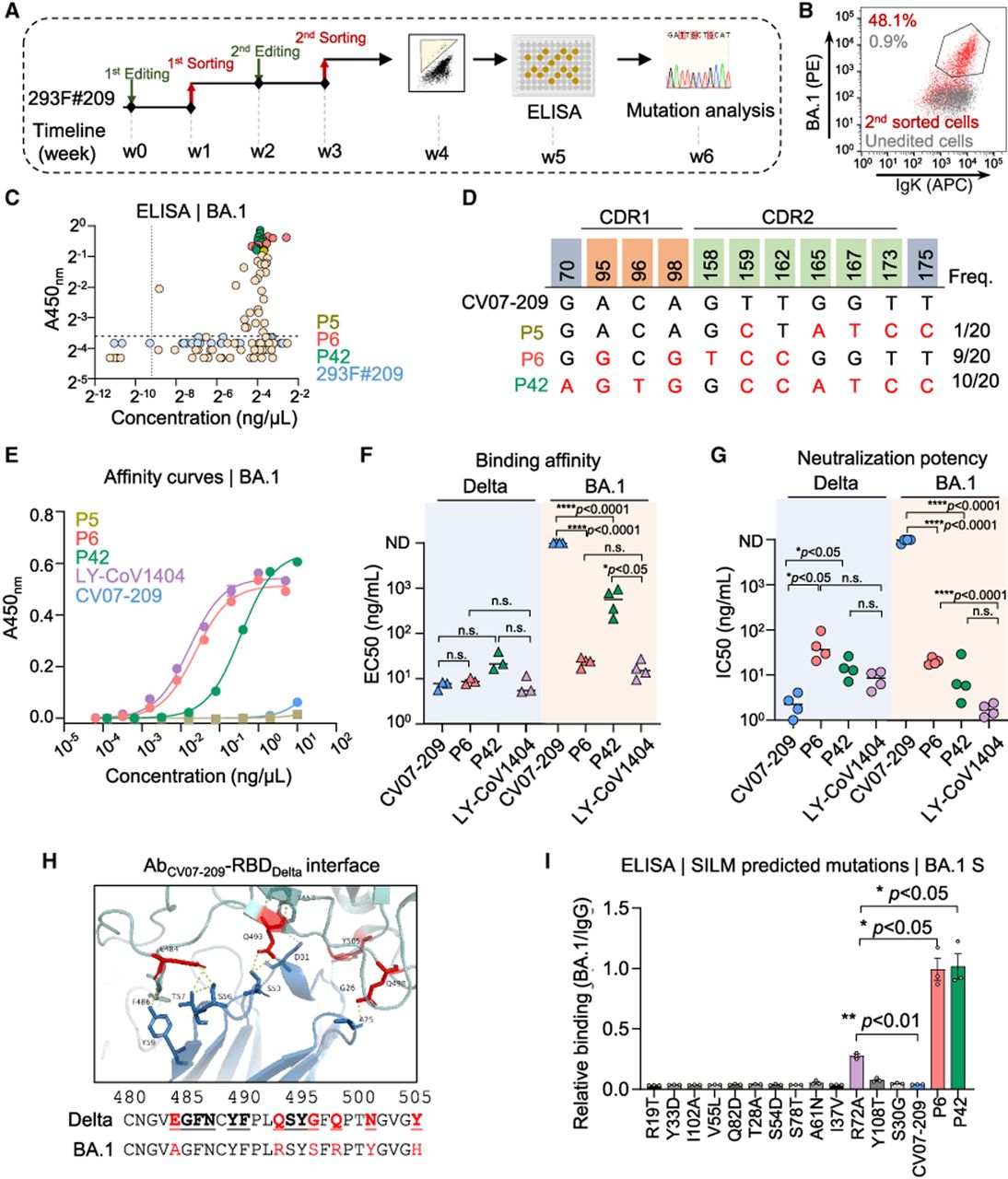
We next expressed and purified the three eAbs to homogeneity for further characterization. LY-CoV1404 (Bebtelovimab), a broadly neutralizing antibody authorized for Omicron treatment, was used as a positive control (Westendorf et al. 2022). ELISA analysis revealed that all three eAbs retained binding affinity toward the Delta S protein. However, only P6 and P42 exhibited robust binding to BA.1 S protein (Fig. 4E,F; Supplemental Fig. S5D). Notably, P6 displayed a binding affinity comparable to LY-CoV1404, demonstrating the efficiency of HAE1-driven evolution toward BA.1 S protein (23.9 ng/mL for P6 vs. 16.8 ng/mL for LY-CoV1404) (Fig. 4F; Supplemental Table S1). Both P6 and P42 not only retained neutralizing activity against the Delta variant but also restored potent activity against BA.1, with extended efficacy toward BA.2 (Fig. 4G; Supplemental Fig. S5E). Of note, although P42 exhibited lower binding affinity compared to P6, it maintained similar neutralizing potency against BA.1, with an IC50 value of 14.4 ng/mL versus 44.6 ng/mL of P6 (Fig. 4G; Supplemental Table S1). Both eAbs showed slightly reduced activity relative to LY-CoV1404, but their evolution from a nonactivated status demonstrated the remarkable capability of HAE1 to rejuvenate the antibody potency against emerging antigenic variants (Fig. 4F,G; Supplemental Table S1).
Recent advances in computational technology have expanded antibody optimization capabilities (Desautels et al. 2024; Hie et al. 2024; Shanker et al. 2024). The structure-informed language model (SILM) has achieved especially remarkable performance in antibody affinity prediction (Graves et al. 2020; Li et al. 2023). We implemented SILM to predicted CV07-209 affinity maturation mutations. Due to the lack of structural information for CV07-209, we initially predicted the structure of CV07-209 and the receptor-binding domain of Delta (Delta-RBD) by AlphaFold 3 (Abramson et al. 2024). The interaction structure revealed an epitope similar to the reported Group B, particularly the 484E-487N region (Fig. 4H). This was evidenced by the findings that the 4-aa mutation form of the Delta S protein disrupted its binding ability to CV07-209 (Supplemental Fig. S5F). Next, we employed the SILM model to predict mutation sites for CV07-209. Among the top 20 predicted hits, 13 mutations were located in the variable region (Fig. 4I). However, only the R72A exhibited a lower level of improvement in affinity compared to P42 and P6, whereas the other predicted ones failed to enhance affinity (Fig. 4I). These results suggest that HAE1 may outperform computational approaches in certain complex antibody evolution scenarios.
Both deaminases in HAE1 promote antibody affinity maturation
The P42 contained three additional amino acid mutations compared to P5, two of which (Y32C and Y33C, CC pair) were shared with P6 (Fig. 4D; Supplemental Fig. S5B). To evaluate the contribution of this CC pair, we performed reverse mutation assays and found that C33 is more critical than C32 in binding and neutralization against BA.1 (Fig. 5A). Specifically, reverting C33 to tyrosine (C33Y) in both P42 and P6 abolished both binding and neutralizing activity against BA.1 but maintained Delta-recognizing activity (Fig. 5B,C; Supplemental Fig. S6A–D). The Y33C mutation was caused by an A > G substitution in the TAC codon, indicating that TadA8e was involved in affinity maturation (Fig. 4D). Similarly, I53S mutation significantly reduced the binding and neutralizing activities of P6 against BA.1 (Fig. 5B; Supplemental Fig. S6A,C). The evolution of S53I involved the AGT > ATC substitution, likely driven by the combined of AIDmono (G > T) and TadA8e (T > C) (Fig. 4D; Supplemental Fig. S1E), further emphasizing the cooperative function of these two deaminases within HAE1. In contrast, the A24T mutation in P42 had only a mild effect on its binding and neutralizing activities (Fig. 5C; Supplemental Fig. S6B,D). These results suggest that the synergy effect between AIDmono and TadA8e broadens the mutation spectrum and enhances antibody potency beyond what could be achieved by AIDmono alone in cultured cells.
Both deaminases in HAE1 facilitate antibody affinity maturation. (A) Amino acid sequences of P6 and P42 with single reverse mutations. Mutations introduced by HAE1 are colored in red and reverse mutations are colored in blue. (B,C) Neutralization potency of reverse mutants of P6 (B) and P42 (C). IC50 values were calculated using GraphPad Prism software for nonlinear regression analysis, with each data point representing an individual biological replicate. Values >10,000 ng/mL or those with unstable fits are considered as nondetectable (ND). Statistical analysis was performed using an unpaired two-tailed t-test, with P values indicated or noted as not significant (n.s.) if P > 0.05. (D) AlphaFold 3-predicted structure of P6 bound to the BA.1 RBD protein. The box shows an enlarged view of the interaction surface between the antibody heavy chain and the RBD. Predicted interacting residuals from RBD are highlighted in yellow, and mutations in the antibodies are colored in red. The potential S protein epitope recognized by CV07-209 is indicated on the bottom box. (E) Relative binding efficiency of antibodies with the indicated epitope mutations. Data are presented as the ratio of mean fluorescence intensity (MFI) of antibody binding (APC) to antigen expression levels (mCherry). Statistical analysis was performed using a paired two-tailed t-test, with results shown as mean ± SEM from three biological replicates. P values are indicated or noted as not significant (n.s.) if P > 0.05.
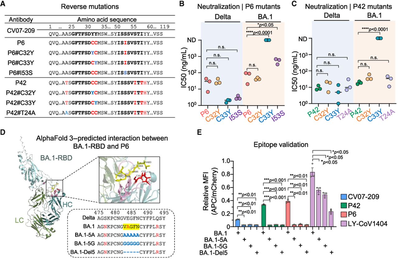
To further characterize the epitope of BA.1 S protein recognized by P6 and P42, we used AlphaFold 3 to predict the interaction surface between the eAbs and the receptor-binding domain (RBD) of BA.1. The prediction suggested a potential interaction between the CC pair and the V483-N487 loop on the BA.1 RBD (Fig. 5D; Supplemental Fig. S6E). This loop is part of the group B epitope previously identified as the binding site for the parent antibody, CV07-209, against wild-type SARS-CoV-2, of which E484A mutation in Omicron may disrupt their interaction (Cao et al. 2022). To confirm this interaction, we mutated the V483-N487 loop into either 5A (all alanine), 5G (all glycine), or deleted it entirely (Del5) (Fig. 5D). The BA.1 S protein and the mutants, fused with mCherry, were expressed on the cell surface and then labeled with eAbs for FACS detection. Disruption of the V483-N487 loop abolished the binding activity of P6 and P42 to BA.1 (Fig. 5E). In contrast, LY-CoV1404, which binds to the group D epitope of the RBD (Cao et al. 2022), retained partial binding to BA.1 with the modified V483-N487 loop (Fig. 5E), highlighting that P6 and P42 potentially interact with the V483-N487 loop of RBD. Our findings suggest that HAE1-driven antibody evolution can rejuvenate binding potency against mutated epitopes.
Discussion
Unlike conventional base editors such as ABEs and CBEs that are designed for precise gene editing to address human diseases caused by point mutations (Rees and Liu 2018), GHM tools require broad mutagenesis capabilities and flexible targeting for antibody diversification as well as other protein engineering. Therefore, HAE1 introduces the PAMless SpRY variant of Cas9 to allow the designed sgRNA targeting any residue within CDRs to enhance mutagenesis potential (Fig. 3A; Supplemental Figs. S1A, S3C). Although the broad targeting range of SpRY poses potential off-target risks that impair precise gene editing, such concerns are less relevant in antibody evolution. Additionally, HAE1 shows a critical advancement by increasing the frequency of A:T mutations (Figs. 1F, 3E), because A:T mutations comprise ∼60% of the total SHM mutations after affinity selection of B cells in vivo (Di Noia and Neuberger 2007; Peled et al. 2008). However, previous ex vivo AID-mediated mutagenesis systems have struggled to generate such mutations efficiently (Martin et al. 2002; Liu et al. 2018; Luo et al. 2020). Although the current HAE1 exhibits restricted A-to-C and A-to-T editing activity, further optimization can be advanced by leveraging recent base-editor designs that integrate the hypoxanthine-excising N-methylpurine DNA glycosylase (MPG) (Tong et al. 2023; Chen et al. 2024), or introducing diverse DNA repair pathways to the host cells to enhance the mutation spectrum. However, the potential cellular toxicity associated with excessively high mutation rates warrants careful evaluation.
Moreover, challenges remain for computational strategies in antibody evolution, particularly due to the need for experimental validation and limitations in computational complexity, which constrain prediction accuracy and the exploration of multiple mutation spaces (Li et al. 2023; Desautels et al. 2024; Erasmus et al. 2024; Shanker et al. 2024). Amplicon sequencing data generated after two-round HAE1-guided mutagenesis provide valuable information on eAbs (Supplemental Fig. S5B). These data can serve as robust, curated data sets for training and improving computational models, thereby enhancing the predictive power of antibody design algorithms (Gao and Skolnick 2024; Buller et al. 2025).
Methods
Plasmid construction
PCR amplification was performed using the PowerPol 2X PCR Mix with Dye (Abclone, #RK20719). Plasmids were constructed by Gibson assembly using the Seamless Cloning kit (Beyotime, #D7010M). Specifically, the sequences encoding AIDmono or TadA8e were cloned and fused to the N terminus of nickase SpRY (D10A) to construct PAMless-AID or HAE1. A BFP reporter gene was coexpressed with a P2A self-cleaving peptide sequence. Additionally, sgRNA plasmids targeting either eGFP or the VH region of CV07-209 were cloned into the sgRNA expression vector under the U6 promoter. All sgRNA sequences are listed in the Supplemental Table S2.
The light and heavy chain sequences of the CV07-209 antibody were synthesized by GENEWIZ and cloned into vectors containing IgK and IL2 secretory signal peptides, respectively, under the control of the EFS and CMV promoters. The constant region of human IgG was also synthesized to facilitate the construction of a full-length human IgG-formatted antibody. Subsequently, the antibody expression cassette was cloned into a pBlue-KS backbone, flanked by an 800-bp homology sequence of AAVSI, to enable precise knock-in through CRISPR-Cas9-mediated homologous recombination. Plasmids used in this study will be deposited to Addgene (https://www.addgene.org, #254231 and #254232), and their complete sequences are provided in Supplemental Table S3.
Cell culture and plasmid transfection
The 293T-eGFP cell line, generated in our previous study for detecting the editing efficiency of different tools (Xin et al. 2022), was used in this work. HEK293F cells (Thermo Fisher Scientific) were used for recombinant antibody expression. Adherent cells were cultured in DMEM (Corning) supplemented with 10% Fetal Bovine Serum (FBS, ExCell Bio), 1% penicillin-streptomycin (Corning), and 1% L-Glutamine (Corning) at 37°C in 5% CO2. Suspended HEK293F cells were cultured on an orbital shaker in Freestyle 293F expression medium (Gibco, #c12338018) at 100 rpm in 8% CO2.
To assess editing efficiency, 293T-eGFP cells were seeded in a 6-well plate and transfected with a mixture of 1.5 µg sgRNA plasmids and 2.5 µg PAMless-AID or HAE1 plasmid by polyethyleneimine (PEI) (Sigma-Aldrich, 1 mg/mL). Approximately 2 × 10⁵ BFP-positive cells were sorted 72 h posttransfection using the Aria SORP flow cytometer, and genomic DNA was extracted for amplicon sequencing preparation.
For antibody evolution, 293F#209 cells were seeded in 6-cm dishes and transfected with a mixture consisting of 4 µg HAE1 or 2 µg AIDmono plasmid and 2 µg plasmid of sgRNA mix targeting CDRs using PEI. Genomic DNA was extracted 72 h posttransfection for amplicon sequencing preparation.
Stable cell line
A stable cell line expressing the CV07-209 antibody was generated through CRISPR-Cas9 mediated transgene knock-in at the AAVS1 safe harbor site in adherent cultured HEK293F cells. In brief, HEK293F cells were cotransfected with a CRISPR-Cas9 vector targeting the AAVS1 locus and a donor plasmid carrying the antibody expression cassette. GFP-positive cells were sorted into 96-well plates 7 days after transfection. Single clones were genotyped by PCR ∼10 days after sorting.
DNA library preparation for amplicon sequencing and data analysis
The targeted GFP loci were PCR-amplified from 500 ng genomic DNA using primers listed in the Supplemental Table S4. An initial PCR of 25 cycles was performed with TranStart Fastpfu high-fidelity DNA polymerase, and a second round of 15 cycles was used to add sequencing adaptor and barcodes. The final PCR products were gel-purified, quantified by DeNovix, and sequenced by PE150 on the NovaSeq 6000 platform (GENEWIZ).
DNA libraries from 293F#209 cells were prepared using an additional liner random barcoded primer extension step with 1–2 µg genomic DNA, following the procedure described above. This process yielded ∼500-bp DNA products, which were gel-purified, quantified by DeNovix, and sequenced by PE300 on the NextSeq 2000 platform (GENEWIZ).
The sequencing data were initially demultiplexed using the FastMultx tool. Paired-end reads were analyzed by CRISPResso2 (Clement et al. 2019). To assess editing efficiency targeting the eGFP locus, a ±15-bp window centered on the midpoint of the 20-bp sgRNA was used. For assessing the editing efficiency in the antibody gene, a ±178-bp window centered on the VH region of the CV07-209 gene was used. All other parameters were set to their default values. Substitution efficiency was calculated from the output file by dividing the number of substitutions by the number of reads aligned. Insertion and deletion efficiencies were calculated using the same method. The distribution of substitutions, deletions, and insertions was analyzed using the file “Nucleotide_percentage_table.txt” and “Modification_count_vectors.txt”. The mutation spectrum was analyzed using the file “Nucleotide_frequency_summary.txt”. For single sgRNA-mediated eGFP editing, only nucleotide conversions within the 20-bp sgRNA-targeted region were considered. For multi-sgRNA-mediated antibody diversification, the total region covered by the sgRNAs was analyzed, spanning 46 bp for CDR1, 34 bp for CDR2, and 58 bp for CDR3.
Antibody sequences from the second round of evolution were subjected to phylogenetic analysis, with sequences occurring fewer than five times excluded. Multiple sequence alignment was performed using MUSCLE (V5.1) (Edgar 2004), and a phylogenetic tree was constructed with FastTree (2.1.11) (Price et al. 2009), rooted with the CV07-209 sequence. The phylogenetic tree was visualized using iTOL.
Protein expression and purification
The DNA sequences of antibody mutants were PCR-amplified and cloned into the antibody expression vector pFuse-hIgG-GFP. A total of 400 mL HEK293F cells, cultured at a concentration of 1 million/mL, were transfected with 400 µg plasmids by PEI. Supernatants containing secreted antibodies were harvested 3.5 days posttransfection and purified using protein G beads (Beyotime, #P2019). Antibody concentration and purity were estimated by SDS-PAGE.
The DNA sequence of SARS-CoV-2 Delta or BA.1 S protein (the extracellular domain, residues 1–1208, with S6P mutation) (Hsieh et al. 2020) with a C-terminal His8 tag was cloned into the pcDNA3.1 vector. Plasmids were transfected into HEK293F cells using PEI at a concentration of 1 million/mL and expressed for 4 days. The S proteins were purified using Ni-NTA resin, followed by size-exclusion chromatography (SEC) on a Superose 6 Increase 10/300 gel filtration column.
S proteins of Delta and BA.1 were biotinylated using EZ-Link Sulfo-NHS-Biotin (Thermo Fisher Scientific, #21331) according to the manufacturer's instructions.
Fluorescence-activated cell sorting–based antibody evolution
Each round of evolution was performed 7 days posttransfection using FACS (BD Aria Fusion). Briefly, 10–20 million cells edited by HAE1 or AIDmono were digested, collected by centrifugation, and washed twice with 1 mL FACS buffer (PBS with 2% FBS). Then, 1 ng/µL biotinylated BA.1 was diluted in 500 µL FACS buffer and resuspended cells for antigen labeling at room temperature (RT) for 25 min. After labeling, the cells were washed twice with FACS buffer and stained with a 1:400 dilution of APC anti-human Ig light chain κ antibody (Biolegend, #TB28-2) and a 1:400 diluted PE-conjugated streptavidin (Sangon Biotech, #D110513) in 200 µL FACS buffer. The cells were incubated in the dark for 15 min, washed twice with FACS buffer, and resuspended with 500 µL FACS buffer for sorting. Sorting gates were set to select cells with improved BA.1 binding, representing 0.5%–1% of the total population. Approximately 1 × 10⁴ to 2 × 10⁴ cells were collected and expanded for the next round of evolution. Single cells from the final round of selection were sorted into 96-well plates and cultured for subsequent identification.
Enzyme-linked immunosorbent assay
To determine antigen binding affinity, Nunc-Immuno MaxiSorp 96-well plates (Thermo Fisher Scientific) were coated with 1 ng/µL of BA.1 diluted in 100 µL PBS and incubated overnight at 4°C. For IgG expression level detection, plates were coated with anti-human IgH/IgL (Biodragon, 2.5 ng/µL) in PBS. After coating, plates were washed three times with PBST (PBS with 0.05% Tween-20) and blocked with 3% BSA in PBST (blocking buffer) for 1 h at RT. Purified antibodies were serially diluted fivefold starting from 10 ng/µL in blocking buffer and then incubated for 2 h at RT. For ELISA with supernatant, 100 µL supernatants from single clones were collected, diluted twofold in blocking buffer, and incubated for 2 h at RT. After three washes with PBST, HRP-conjugated anti-human IgH/L (Proteintech, #SA00001-17) diluted 1:10,000 was added and incubated for 1 h at RT. Following the final three washes with PBST, 100 µL TMB Substrate Solution (Beyotime, #P0209) was added for the HRP reaction for 10 min, and the reaction was stopped with 100 µL Stop Solution (Beyotime, #P0215). Absorbance was measured at 450 nm using BioTek Cytation5. Data were analyzed by GraphPad prism, and the curves were fitted with a four-parameter nonlinear regression model to determine the EC50 values.
Pseudovirus neutralization assay
A pseudovirus neutralization assay was conducted to evaluate the neutralizing activity of antibodies. Generally, 293T-ACE2 cells (4 × 10⁴ cells per well) were seeded in 96-well plates 24 h before infection. Serially diluted antibodies were incubated with an equal volume of virus for 1 h at 37°C. The antibody-virus mixture was then added to the 293T-ACE2 cells. Luciferase efficiency was measured 48 h later by adding D-luciferin substrate (D-Luciferin potassium salt, Beyotime, #ST196) into the cells, and luminescence intensity was detected by BioTek Cytation5. Data were analyzed using GraphPad prism software, and the curves were fitted with a four-parameter nonlinear regression model to calculate IC50 (half maximal inhibitory concentration). Each biological replicate consisted of three technical replicates, and at least three biological repeats were conducted to determine the IC50.
Prediction of antibody structure and affinity maturation mutations
The three-dimensional structures of the antibodies and the S protein were predicted using AlphaFold 3 (online version of AlphaFold Server). Specifically, we focused on the RBD of the S protein and the Fab region of antibody, as these regions play a crucial role in the antibody-antigen interactions. The amino acid sequences of the RBD, heavy chain and light chain of CV07-209 Fab are listed in the Supplemental Table S5.
The SILM model was employed for predicting affinity maturation mutations. Briefly, the SILM model was installed following the instructions provided on GitHub (https://github.com/varun-shanker/structural-evolution). The interaction structure file generated by AlphaFold 3 (CV07-209 and Delta-RBD) was used as input for the prediction, with default parameters applied. In this study, only the predictions for the heavy chain were analyzed.
Statistical analysis
All statistical analyses were conducted using GraphPad Prism 9. The number of replicates and the specific statistical tests used are detailed in the corresponding figure legends. Data are presented as mean ± SEM, and statistical significance was defined as P < 0.05.
Data access
All raw and processed sequencing data generated in this study have been submitted to the NCBI Gene Expression Omnibus (GEO; https://www.ncbi.nlm.nih.gov/geo/) under accession numbers GSE281292 and GSE281195.
Competing interest statement
J.H., Y.W., and Y.G. have filed a patent application related to the work described here. The title of the patent application is “A base editor and its application.” China Provisional Application was filed on 11/26/2025 (CN202511756586.9). The remaining authors declare no competing interests.
Acknowledgments
We thank Drs. Xiaohui Zhang and Dali Li for their gifts of A&CBEmax plasmids. We thank the Flow Cytometry Core at the National Center for Protein Sciences, Peking University, particularly Xuefang Zhang, Yabin Bian, and Hui Li, for their technical help. This work is supported by the National Key Research & Development Program of China (2022YFC3400201), the National Natural Science Foundation of China grant (32425015), the Ministry of Agriculture and Rural Affairs of China and the Sichuan Provincial Natural Science Foundation (2025ZNSFSC0236). J.H. is an investigator at the PKU-TSU Center for Life Sciences.
Author contributions: Y.W., Y.G., and J.H. conceived and designed the research; Y.W., H.X., T.G., and R.L. constructed the plasmids and cell lines; Y.W. and Y.G. performed high-throughput sequencing; F.-L.M. contributed to experimental design and provided the original plasmids; Y.W., X.L., and Y.G. analyzed the sequencing data; Y.W., H.X., J.C., Q.L., R.P., S.Y., and Z.Y. prepared cell samples and purified antibodies; Y.W., H.X., J.C., Q.L., and R.L. performed ELISA and peudovirus assay; Y.W., Y.G., and J.H. wrote the manuscript.
Notes
[1] Supplementary material [Supplemental material is available for this article.]
[2] Article published online before print. Article, supplemental material, and publication date are at https://www.genome.org/cgi/doi/10.1101/gr.281396.125.
References
- ↵Abramson J, Adler J, Dunger J, Evans R, Green T, Pritzel A, Ronneberger O, Willmore L, Ballard AJ, Bambrick J, 2024. Accurate structure prediction of biomolecular interactions with AlphaFold 3. Nature 630: 493–500. 10.1038/s41586-024-07487-w
- ↵Alt FW, Zhang Y, Meng FL, Guo C, Schwer B. 2013. Mechanisms of programmed DNA lesions and genomic instability in the immune system. Cell 152: 417–429. 10.1016/j.cell.2013.01.007
- ↵Bai X, Xu L, Wang Z, Zhuang X, Ning J, Sun Y, Wang H, Guo Y, Xu Y, Guo J, 2025. Computational-aided rational mutation design of pertuzumab to overcome active HER2 mutation S310F through antibody–drug conjugates. Proc Natl Acad Sci 122: 2017. 10.1073/pnas.2413686122
- ↵Bowers PM, Horlick RA, Neben TY, Toobian RM, Tomlinson GL, Dalton JL, Jones HA, Chen A, Altobell L, Zhang X, 2011. Coupling mammalian cell surface display with somatic hypermutation for the discovery and maturation of human antibodies. Proc Natl Acad Sci 108: 20455–20460. 10.1073/pnas.1114010108
- ↵Briney B, Inderbitzin A, Joyce C, Burton DR. 2019. Commonality despite exceptional diversity in the baseline human antibody repertoire. Nature 566: 393–397. 10.1038/s41586-019-0879-y
- ↵Buller R, Damborsky J, Hilvert D, Bornscheuer UT. 2025. Structure prediction and computational protein design for efficient biocatalysts and bioactive proteins. Angew Chemie Int Ed 64: e202421686. 10.1002/anie.202421686
- ↵Cao Y, Wang J, Jian F, Xiao T, Song W, Yisimayi A, Huang W, Li Q, Wang P, An R, 2022. Omicron escapes the majority of existing SARS-CoV-2 neutralizing antibodies. Nature 602: 657–663. 10.1038/s41586-021-04385-3
- ↵Carter PJ, Rajpal A. 2022. Designing antibodies as therapeutics. Cell 185: 2789–2805. 10.1016/j.cell.2022.05.029
- ↵Chao G, Lau WL, Hackel BJ, Sazinsky SL, Lippow SM, Wittrup KD. 2006. Isolating and engineering human antibodies using yeast surface display. Nat Protoc 1: 755–768. 10.1038/nprot.2006.94
- ↵Chen S, Qiu J, Chen C, Liu C, Liu Y, An L, Jia J, Tang J, Wu L, Hang H. 2012. Affinity maturation of anti-TNF-alpha scFv with somatic hypermutation in non-B cells. Protein Cell 3: 460–469. 10.1007/s13238-012-2024-7
- ↵Chen L, Hong M, Luan C, Gao H, Ru G, Guo X, Zhang D, Zhang S, Li C, Wu J, 2024. Adenine transversion editors enable precise, efficient A•T-to-C•G base editing in mammalian cells and embryos. Nat Biotechnol 42: 638–650. 10.1038/s41587-023-01821-9
- ↵Clement K, Rees H, Canver MC, Gehrke JM, Farouni R, Hsu JY, Cole MA, Liu DR, Joung JK, Bauer DE, 2019. CRISPResso2 provides accurate and rapid genome editing sequence analysis. Nat Biotechnol 37: 224–226. 10.1038/s41587-019-0032-3
- ↵Cumbers SJ, Williams GT, Davies SL, Grenfell RL, Takeda S, Batista FD, Sale JE, Neuberger MS. 2002. Generation and iterative affinity maturation of antibodies in vitro using hypermutating B-cell lines. Nat Biotechnol 20: 1129–1134. 10.1038/nbt752
- ↵Desautels TA, Arrildt KT, Zemla AT, Lau EY, Zhu F, Ricci D, Cronin S, Zost SJ, Binshtein E, Scheaffer SM, 2024. Computationally restoring the potency of a clinical antibody against Omicron. Nature 629: 878–885. 10.1038/s41586-024-07385-1
- ↵Devilder MC, Moyon M, Gautreau-Rolland L, Navet B, Perroteau J, Delbos F, Gesnel MC, Breathnach R, Saulquin X. 2019. Ex vivo evolution of human antibodies by CRISPR-X: from a naive B cell repertoire to affinity matured antibodies. BMC Biotechnol 19: 14. 10.1186/s12896-019-0504-z
- ↵Di Noia JM, Neuberger MS. 2007. Molecular mechanisms of antibody somatic hypermutation. Annu Rev Biochem 76: 1–22. 10.1146/annurev.biochem.76.061705.090740
- ↵Edgar RC. 2004. MUSCLE: multiple sequence alignment with high accuracy and high throughput. Nucleic Acids Res 32: 1792–1797. 10.1093/nar/gkh340
- ↵Erasmus MF, Spector L, Ferrara F, DiNiro R, Pohl TJ, Perea-Schmittle K, Wang W, Tessier PM, Richardson C, Turner L, 2024. AIntibody: an experimentally validated in silico antibody discovery design challenge. Nat Biotechnol 42: 1637–1642. 10.1038/s41587-024-02469-9
- ↵Gao M, Skolnick J. 2024. Improved deep learning prediction of antigen–antibody interactions. Proc Natl Acad Sci 121: 2017. 10.1073/pnas.2410529121
- ↵Gaudelli NM, Komor AC, Rees HA, Packer MS, Badran AH, Bryson DI, Liu DR. 2017. Programmable base editing of A•T to G•C in genomic DNA without DNA cleavage. Nature 551: 464–471. 10.1038/nature24644
- ↵Graves J, Byerly J, Priego E, Makkapati N, Parish S, Medellin B, Berrondo M. 2020. A review of deep learning methods for antibodies. Antibodies 9: 12. 10.3390/antib9020012
- ↵Grünewald J, Zhou R, Lareau CA, Garcia SP, Iyer S, Miller BR, Langner LM, Hsu JY, Aryee MJ, Joung JK. 2020. A dual-deaminase CRISPR base editor enables concurrent adenine and cytosine editing. Nat Biotechnol 38: 861–864. 10.1038/s41587-020-0535-y
- ↵Guzzardo PM, Rashkova C, Dos Santos RL, Tehrani R, Collin P, Bürckstümmer T. 2017. A small cassette enables conditional gene inactivation by CRISPR/Cas9. Sci Rep 7: 16770. 10.1038/s41598-017-16931-z
- ↵Hao Q, Zhan C, Lian C, Luo S, Cao W, Wang B, Xie X, Ye X, Gui T, Voena C, 2023. DNA repair mechanisms that promote insertion-deletion events during immunoglobulin gene diversification. Sci Immunol 8: eade1167. 10.1126/sciimmunol.ade1167
- ↵Hawkins RE, Russell SJ, Winter G. 1992. Selection of phage antibodies by binding affinity: mimicking affinity maturation. J Mol Biol 226: 889–896. 10.1016/0022-2836(92)90639-2
- ↵He W, Ou T, Skamangas N, Bailey CC, Bronkema N, Guo Y, Yin Y, Kobzarenko V, Zhang X, Pan A, 2023. Heavy-chain CDR3-engineered B cells facilitate in vivo evaluation of HIV-1 vaccine candidates. Immunity 56: 2408–2424.e6. 10.1016/j.immuni.2023.07.003
- ↵Hegde PS, Chen DS. 2020. Top 10 challenges in cancer immunotherapy. Immunity 52: 17–35. 10.1016/j.immuni.2019.12.011
- ↵Hendel SJ, Shoulders MD. 2021. Directed evolution in mammalian cells. Nat Methods 18: 346–357. 10.1038/s41592-021-01090-x
- ↵Hess GT, Frésard L, Han K, Lee CH, Li A, Cimprich KA, Montgomery SB, Bassik MC. 2016. Directed evolution using dCas9-targeted somatic hypermutation in mammalian cells. Nat Methods 13: 1036–1042. 10.1038/nmeth.4038
- ↵Hess GT, Tycko J, Yao D, Bassik MC. 2017. Methods and applications of CRISPR-mediated base editing in eukaryotic genomes. Mol Cell 68: 26–43. 10.1016/j.molcel.2017.09.029
- ↵Hie BL, Shanker VR, Xu D, Bruun TUJ, Weidenbacher PA, Tang S, Wu W, Pak JE, Kim PS. 2024. Efficient evolution of human antibodies from general protein language models. Nat Biotechnol 42: 275–283. 10.1038/s41587-023-01763-2
- ↵Hsieh CL, Goldsmith JA, Schaub JM, DiVenere AM, Kuo HC, Javanmardi K, Le KC, Wrapp D, Lee AG, Liu Y, 2020. Structure-based design of prefusion-stabilized SARS-CoV-2 spikes. Science 369: 1501–1505. 10.1126/science.abd0826
- ↵Komor AC, Kim YB, Packer MS, Zuris JA, Liu DR. 2016. Programmable editing of a target base in genomic DNA without double-stranded DNA cleavage. Nature 533: 420–424. 10.1038/nature17946
- ↵Kreye J, Reincke SM, Kornau HC, Sánchez-Sendin E, Corman VM, Liu H, Yuan M, Wu NC, Zhu X, Lee CCD, 2020. A therapeutic non-self-reactive SARS-CoV-2 antibody protects from lung pathology in a COVID-19 hamster model. Cell 183: 1058–1069.e19. 10.1016/j.cell.2020.09.049
- ↵Kuramochi T, Gan SW, Ho AWS, Wang B, Kageji N, Nambu T, Iida S, Okuda-Miura M, Chia WS, Yeo CY, 2022. Comprehensive engineering of a therapeutic neutralizing antibody targeting SARS-CoV-2 spike protein to neutralize escape variants. MAbs 14: 2040350. 10.1080/19420862.2022.2040350
- ↵Li J, Kang G, Wang J, Yuan H, Wu Y, Meng S, Wang P, Zhang M, Wang Y, Feng Y, 2023. Affinity maturation of antibody fragments: a review encompassing the development from random approaches to computational rational optimization. Int J Biol Macromol 247: 125733. 10.1016/j.ijbiomac.2023.125733
- ↵Liang Y, Xie J, Zhang Q, Wang X, Gou S, Lin L, Chen T, Ge W, Zhuang Z, Lian M, 2022. AGBE: a dual deaminase-mediated base editor by fusing CGBE with ABE for creating a saturated mutant population with multiple editing patterns. Nucleic Acids Res 50: 5384–5399. 10.1093/nar/gkac353
- ↵Lin H, Yang X, Ye S, Huang L, Mu W, Lin H, Yang X, Ye S, Huang L, Mu W, 2024. Antigen escape in CAR-T cell therapy: mechanisms and overcoming strategies. Biomed Pharmacother 178: 117252. 10.1016/j.biopha.2024.117252
- ↵Liu LD, Huang M, Dai P, Liu T, Fan S, Cheng X, Zhao Y, Yeap LS, Meng FL. 2018. Intrinsic nucleotide preference of diversifying base editors guides antibody ex vivo affinity maturation. Cell Rep 25: 884–892.e3. 10.1016/j.celrep.2018.09.090
- ↵Luo R, Zhao Y, Fan Y, An L, Jiang T, Ma S, Hang H. 2020. High efficiency CHO cell display-based antibody maturation. Sci Rep 10: 8102. 10.1038/s41598-020-65044-7
- ↵Manso T, Folch G, Giudicelli V, Jabado-Michaloud J, Kushwaha A, Nguefack Ngoune V, Georga M, Papadaki A, Debbagh C, Pégorier P, 2022. IMGT databases, related tools and web resources through three main axes of research and development. Nucleic Acids Res 50: D1262–D1272. 10.1093/nar/gkab1136
- ↵Martin A, Scharff MD. 2002. Somatic hypermutation of the AID transgene in B and non-B cells. Proc Natl Acad Sci 99: 12304–12308. 10.1073/pnas.192442899
- ↵Martin A, Bardwell PD, Woo CJ, Fan M, Shulman MJ, Scharff MD. 2002. Activation-induced cytidine deaminase turns on somatic hypermutation in hybridomas. Nature 415: 802–806. 10.1038/nature714
- ↵Masuda H, Sawada A, Hashimoto SI, Tamai K, Lin KY, Harigai N, Kurosawa K, Ohta K, Seo H, Itou H. 2022. Fast-tracking antibody maturation using a B cell-based display system. MAbs 14: 2122275. 10.1080/19420862.2022.2122275
- ↵Molina RS, Rix G, Mengiste AA, Álvarez B, Seo D, Chen H, Hurtado JE, Zhang Q, García-García JD, Heins ZJ, 2022. In vivo hypermutation and continuous evolution. Nat Rev Methods Prim 2: 36. 10.1038/s43586-022-00119-5
- ↵Muecksch F, Weisblum Y, Barnes CO, Schmidt F, Schaefer-Babajew D, Wang Z, Julio JC, Flyak AI, DeLaitsch AT, Huey-Tubman KE, 2021. Affinity maturation of SARS-CoV-2 neutralizing antibodies confers potency, breadth, and resilience to viral escape mutations. Immunity 54: 1853–1868.e7. 10.1016/j.immuni.2021.07.008
- ↵Mullard A. 2021. FDA approves 100th monoclonal antibody product. Nat Rev Drug Discov 20: 491–495. 10.1038/d41573-021-00079-7
- ↵Nahmad AD, Lazzarotto CR, Zelikson N, Kustin T, Tenuta M, Huang D, Reuveni I, Nataf D, Raviv Y, Horovitz-Fried M, 2022. In vivo engineered B cells secrete high titers of broadly neutralizing anti-HIV antibodies in mice. Nat Biotechnol 40: 1241–1249. 10.1038/s41587-022-01328-9
- ↵Nie J, Huang W, Liu Q, Wang Y. 2020. HIV-1 pseudoviruses constructed in China regulatory laboratory. Emerg Microbes Infect 9: 32–41. 10.1080/22221751.2019.1702479
- ↵Oostindie SC, Lazar GA, Schuurman J, Parren PWHI. 2022. Avidity in antibody effector functions and biotherapeutic drug design. Nat Rev Drug Discov 21: 715–735. 10.1038/s41573-022-00501-8
- ↵Paul S, Konig MF, Pardoll DM, Bettegowda C, Papadopoulos N, Wright KM, Gabelli SB, Ho M, van Elsas A, Zhou S. 2024. Cancer therapy with antibodies. Nat Rev Cancer 24: 399–426. 10.1038/s41568-024-00690-x
- ↵Peled JU, Fei LK, Iglesias-Ussel MD, Roa S, Kalis SL, Goodman MF, Scharff MD. 2008. The biochemistry of somatic hypermutation. Annu Rev Immunol 26: 481–511. 10.1146/annurev.immunol.26.021607.090236
- ↵Peled S, Guez-Haddad J, Zur Biton N, Nimrod G, Fischman S, Fastman Y, Ofran Y. 2024. Computational engineering of a therapeutic antibody to inhibit multiple mutants of HER2 without compromising inhibition of the canonical HER2. eLife 13: RP89814. 10.7554/eLife.89814.2
- ↵Price MN, Dehal PS, Arkin AP. 2009. FastTree: computing large minimum evolution trees with profiles instead of a distance matrix. Mol Biol Evol 26: 1641–1650. 10.1093/molbev/msp077
- ↵Qiao Q, Wang L, Meng FL, Hwang JK, Alt FW, Wu H. 2017. AID recognizes structured DNA for class switch recombination. Mol Cell 67: 361–373.e4. 10.1016/j.molcel.2017.06.034
- ↵Raybould MIJ, Kovaltsuk A, Marks C, Deane CM. 2021. CoV-AbDab: the coronavirus antibody database. Bioinformatics 37: 734–735. 10.1093/bioinformatics/btaa739
- ↵Rees HA, Liu DR. 2018. Base editing: precision chemistry on the genome and transcriptome of living cells. Nat Rev Genet 19: 770–788. 10.1038/s41576-018-0059-1
- ↵Richter MF, Zhao KT, Eton E, Lapinaite A, Newby GA, Thuronyi BW, Wilson C, Koblan LW, Zeng J, Bauer DE, 2020. Phage-assisted evolution of an adenine base editor with improved Cas domain compatibility and activity. Nat Biotechnol 38: 901. 10.1038/s41587-020-0562-8
- ↵Shanker VR, Bruun TUJ, Hie BL, Kim PS. 2024. Unsupervised evolution of protein and antibody complexes with a structure-informed language model. Science 385: 46–53. 10.1126/science.adk8946
- ↵Taylor PC, Adams AC, Hufford MM, de la Torre I, Winthrop K, Gottlieb RL. 2021. Neutralizing monoclonal antibodies for treatment of COVID-19. Nat Rev Immunol 21: 382–393. 10.1038/s41577-021-00542-x
- ↵Tong H, Wang X, Liu Y, Liu N, Li Y, Luo J, Ma Q, Wu D, Li J, Xu C, 2023. Programmable A-to-Y base editing by fusing an adenine base editor with an N-methylpurine DNA glycosylase. Nat Biotechnol 41: 1080–1084. 10.1038/s41587-022-01595-6
- ↵Victora GD, Nussenzweig MC. 2012. Germinal centers. Annu Rev Immunol 30: 429–457. 10.1146/annurev-immunol-020711-075032
- ↵Voss JE, Gonzalez-Martin A, Andrabi R, Fuller RP, Murrell B, McCoy LE, Porter K, Huang D, Li W, Sok D, 2019. Reprogramming the antigen specificity of B cells using genome-editing technologies. eLife 8: e42995. 10.7554/eLife.42995
- ↵Walton RT, Christie KA, Whittaker MN, Kleinstiver BP. 2020. Unconstrained genome targeting with near-PAMless engineered CRISPR-Cas9 variants. Science 368: 290–296. 10.1126/science.aba8853
- ↵Wang Y, Zhang S, Yang X, Hwang JK, Zhan C, Lian C, Wang C, Gui T, Wang B, Xie X, 2023. Mesoscale DNA feature in antibody-coding sequence facilitates somatic hypermutation. Cell 186: 2193–2207.e19. 10.1016/j.cell.2023.03.030
- ↵Westendorf K, Žentelis S, Wang L, Foster D, Vaillancourt P, Wiggin M, Lovett E, van der Lee R, Hendle J, Pustilnik A, 2022. LY-CoV1404 (bebtelovimab) potently neutralizes SARS-CoV-2 variants. Cell Rep 39: 110812. 10.1016/j.celrep.2022.110812
- ↵Wu Y, Wang F, Shen C, Peng W, Li D, Zhao C, Li Z, Li S, Bi Y, Yang Y, 2020. A noncompeting pair of human neutralizing antibodies block COVID-19 virus binding to its receptor ACE2. Science 368: 1274–1278. 10.1126/science.abc2241
- ↵Xin C, Yin J, Yuan S, Ou L, Liu M, Zhang W, Hu J. 2022. Comprehensive assessment of miniature CRISPR-Cas12f nucleases for gene disruption. Nat Commun 13: 5623. 10.1038/s41467-022-33346-1
- ↵Yeap LS, Hwang JK, Du Z, Meyers RM, Meng FL, Jakubauskaite A, Liu M, Mani V, Neuberg D, Kepler TB, 2015. Sequence-intrinsic mechanisms that target AID mutational outcomes on antibody genes. Cell 163: 1124–1137. 10.1016/j.cell.2015.10.042
- ↵Yin Y, Guo Y, Jiang Y, Quinlan B, Peng H, Crynen G, He W, Zhang L, Ou T, Bailey CC, 2024. In vivo affinity maturation of mouse B cells reprogrammed to express human antibodies. Nat Biomed Eng 8: 361–379. 10.1038/s41551-024-01179-6
- ↵Zhang X, Zhu B, Chen L, Xie L, Yu W, Wang Y, Li L, Yin S, Yang L, Hu H, 2020. Dual base editor catalyzes both cytosine and adenine base conversions in human cells. Nat Biotechnol 38: 856–860. 10.1038/s41587-020-0527-y