
In Silico Cloning of Novel Endothelial-Specific Genes
Abstract
The endothelium plays a pivotal role in many physiological and pathological processes and is known to be an exceptionally active transcriptional site. To advance our understanding of endothelial cell biology and to elucidate potential pharmaceutical targets, we developed a new database screening approach to permit identification of novel endothelial-specific genes. The UniGene gene index was screened using high stringency BLAST against a pool of endothelial expressed sequence tags (ESTs) and a pool of nonendothelial ESTs constructed from cell-type–specific dbEST libraries. UniGene clusters with matches in the endothelial pool and no matches in the nonendothelial pool were selected. The UniGene/EST approach was then combined with serial analysis of gene expression (SAGE) library subtraction and reverse transcription polymerase chain reaction to further examine interesting clusters. Four novel genes were identified and labeled: endothelial cell–specific molecules (ECSM) 1–3 and magic roundabout (similar to the axon guidance protein roundabout). In summary, we present a powerful novel approach for comparative expression analysis combining two datamining strategies followed by experimental verification.
In the postgenomic era, data analysis rather than data collection will present the biggest challenge to biologists. Efforts to ascribe biological meaning to genomic data, whether by identification of function, structure, or expression pattern, are lagging behind sequencing efforts (Boguski 1999). Here, we describe the use of two independent strategies for differential expression analysis combined with experimental verification to identify genes specifically or preferentially expressed in vascular endothelium.
The first strategy was based on an EST cluster expression analysis in the human UniGene gene index (Schuler et al. 1997). Recurrent gappedBLAST searches (Altschul et al. 1997) were performed at very high stringency against expressed sequence tags (ESTs) grouped into two pools. The two pools comprised endothelial cell and nonendothelial cell libraries derived from dbEST (Boguski et al. 1995). The second strategy used another datamining tool: SAGEmapxProfiler. xProfiler is a freely available online tool, which is a part of the NCBI's Cancer Genome Anatomy Project (CGAP) (Cole et al. 1995; Strausberg et al. 1997).
These two approaches alone produced a discouragingly high number of false positives. However, when both strategies were combined, predictions proved exceptionally reliable and four novel candidate endothelial-specific genes have been identified. For two of these genes, full-length cDNAs have been identified in sequence databases. Another gene (EST cluster) corresponds to a partial cDNA sequence from a large-scale cDNA sequencing project and contains a region of similarity to the intracellular domain of human roundabout homolog 1 (ROBO1).
RESULTS
UniGene/EST Gene Index Screen
A pool of endothelial ESTs and a pool of nonendothelial ESTs were extracted using the Sequence Retrieval System (SRS) from dbEST. The endothelial pool consisted of 11,117 ESTs from nine human endothelial libraries (Table 1). The nonendothelial pool included 173,137 ESTs from 108 human cell lines and microdissected tumor libraries (Table 2). ESTs were extracted from dbEST, release April 2000. Multiple-FASTA files were transformed into BLAST searchable databases using the pressdb program. Table 3 shows the expression status of five known endothelial cell–specific genes in these two pools: von Willebrand factor (vWF; Ginsburg et al. 1985); two vascular endothelial growth factor receptors, fms-like tyrosine kinase 1 (FLT1; Shibuya et al. 1990) and kinase insert domain receptor (KDR; Matthews et al. 1991); tyrosine kinase receptor type tie (TIE1; Partanen et al. 1992); and tyrosine kinase receptor type tek (TIE2/TEK; Vikkula et al. 1996).
Nine Human Endothelial Libraries from dbEST
Nonendothelial dbEST Libraries
Five Known Endothelial-Specific Genes in the dbEST Pools
Optimizing the BLAST E-value was crucial for the success of BLAST identity-level searches. Too high an E-value would result in gene paralogs being reported. In contrast, too low (stringent) an E-parameter would result in many false negatives, i.e., true positives would not be reported because of sequencing errors in EST data; ESTs are large-scale, low-cost single pass sequences and have a high error rate (Aaronson et al. 1996). In this work an E-value of 10e–20 was used in searches against the nonendothelial EST pool and a more stringent 10e–30 value was used in searches against the smaller endothelial pool. These values were deemed optimal after a series of test BLAST searches.
SAGE Data and SAGEmap xProfiler Differential Analysis
Internet-based SAGE library subtraction (SAGEmapxProfiler) was used as the second datamining strategy for the identification of novel endothelial-specific or preferentially endothelial genes. Two endothelial SAGE libraries (SAGE_Duke_HMVEC and SAGE_Duke_HMVEC + VEGF with a total of 110,790 sequences) were compared with 24 nonendothelial cell line libraries (full list in Table4, total of 733,461 sequences). Table5 shows the status of expression of the five reference endothelial-specific genes in these two SAGE pools.
Twenty-four Nonendothelial Cell Serial Analysis Gene Expression (SAGE)–Cancer Genome Anatomy Project
Five Known Endothelial-Specific Genes in the CGAP SAGE Pools
Combined Data Gives Highly Accurate Predictions
Twenty known genes were selected in the UniGene/EST screen (Table6). These genes had no matches in the nonendothelial pool and at least one match in the endothelial pool. The list contained four endothelial-specific genes: TIE1 (Partanen et al. 1992), TIE2/TEK (Vikkula et al. 1996), LYVE1(Banerji et al. 1999), and multimerin (Hayward et al. 1998), indicating ∼20% accuracy of prediction. Other genes on the list, although certainly preferentially expressed in the endothelial cells, may not be endothelial specific. To improve on the prediction accuracy, we decided to combine UniGene/EST screen with thexProfiler SAGE analysis. Table7 shows how data from the two approaches were combined. Identity-level BLAST searches were performed on mRNAs (known genes) or phrap-computed contigs (EST clusters representing novel genes) to investigate how these genes were represented in the endothelial and nonendothelial pool. Subsequent experimental verification by reverse transcription polymerase chain reaction (RT-PCR; Fig. 1) proved that the combined approach was 100% accurate, i.e., genes on the xProfilerlist that had no matches the nonendothelial EST pool and at least one match in the endothelial pool were indeed endothelial specific.
Results of the UniGene/EST Screen
xProfiler Differential Analysis was Combined with Data from the UniGene/EST Screen Achieving 100% Certainty of Prediction
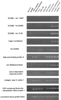
Experimental verification by reverse transcription polymerase chain reaction (RT-PCR). Candidate endothelial-specific genes predicted by the combination of the UniGene/EST screen and xProfilerserial analysis of gene expression (SAGE) differential analysis (Table8) were checked for expression in three endothelial and nine nonendothelial cell cultures. Endothelial cultures were as follows: HMVEC (human microvascular endothelial cells), HUVEC (human umbilical vein endothelial cells) confluent culture, and HUVEC proliferating culture. Nonendothelial cultures were as follows: normal endometrial stromal (NES) cells grown in normoxia and NES grown in hypoxia, MDA 453 and MDA 468 breast carcinoma cell lines, HeLa, FEK4 fibroblasts cultured in normoxia and FEK4 fibroblasts cultured in hypoxia, SW480, and HCT116, the last two being colorectal epithelium cell lines.ECSM1 and ECSM2 showed complete endothelial specificity, whereas ECSM3 and magic roundabout were very strongly preferentially expressed in the endothelium. Interestingly, all these novel genes appear more specific than the benchmark endothelial-specific gene, von Willebrand factor.
DISCUSSION
There have been several reports of computer analysis of tissue transcriptosomes. Usually an expression profile is constructed, based on the number of tags assigned to a given gene or a class of genes (Bernstein et al. 1996; Welle et al. 1999; Bortoluzzi et al. 2000). An attempt can be made to identify tissue-specific transcripts: for example, Vasmatzis et al. (1997) described three novel genes expressed exclusively in the prostate by in silico subtraction of libraries from the dbEST collection. Purpose-made cDNA libraries may also be used. Ten candidate granulocyte-specific genes have been identified by extensive sequence analysis of cDNA libraries derived from granulocytes and eleven other tissue samples, namely a hepatocyte cell line, fetal liver, infant liver, adult liver, subcutaneous fat, visceral fat, lung, colonic mucosa, keratinocytes, cornea, and retina (Itoh et al. 1998).
An analysis similar to the dbEST-based approach taken by Vasmatzis et al. (1997) is complicated by the fact that endothelial cells are present in all tissues of the body and endothelial ESTs are contaminating all bulk tissue libraries. To validate this, we used three well-known endothelial-specific genes—KDR,FLT1, and TIE-2—as queries for BLAST searches against dbEST. Transcripts were present in a wide range of tissues, with multiple hits in well-vascularized tissues (e.g., placenta, retina), embryonic tissues (liver, spleen), or infant tissues (brain). In addition, we found that simple subtraction of endothelial EST libraries against all other dbEST libraries failed to identify any specific genes (data not shown).
Two very different types of expression data resources were used in our datamining efforts. The UniGene/EST screen was based on EST libraries from dbEST. There are nine human endothelial libraries in the current release of dbEST, with a relatively small total number of ESTs (11,117). Some well-known endothelial-specific genes are not represented in this data set (Table 3). This limitation raised our concerns that genes with low levels of expression would be overlooked in our analysis. Therefore, we used another type of computable expression data: CGAP SAGE libraries. SAGE tags are sometimes called small ESTs (usually 10–11 bp in length). Their major advantage is that they can be unambiguously located within the cDNA: they are immediately adjacent to the most 3′ NlaIII restriction site. Although there are only two endothelial CGAP SAGE libraries available at the moment, they contain an impressive total of ∼111,000 tags—a data set approximately ten times bigger than the 11,117 sequences in the endothelial EST pool. The combined approach proved very accurate (Fig.1; Table 8) when verified by RT-PCR. We report here identification of four novel highly endothelial-specific genes: endothelial cell–specific molecule 1 (ECSM1; UniGene entry Hs.13957), endothelial cell–specific molecule 2 (ECSM2; UniGene entry Hs.30089), endothelial cell–specific molecule 3 (ECSM3; UniGene entry Hs.8135), and magic roundabout (UniGene entry Hs.111518). For a comprehensive summary of data available on these genes, see Table 8.
Summary of Available Information on ECSM1-3 and Magic Roundabout
ECSM1 has no protein or nucleotide homologs. It codes for a small protein of ∼103 aa, the longest and most upstream open reading frame (ORF) identified in the contig sequence.
BLAST searches against the EMBL patent database revealed thatECSM2 corresponds to the cDNA from the patent “cDNA encoding novel polypeptide from human umbilical vein endothelial cell” (Shibayama et al. 1997), EMBL acc. E10591. A 205-aa polypeptide coded by this cDNA is a transmembrane protein with a suggested role in cell adhesion in that it is serine and proline rich, although no exact function has yet been identified.
ECSM3 was found to be identical with the matrix remodeling–associated gene 4 (MXRA4, cDNA sequence acc.AW888224) recently identified in a screen of 40,000 genes from 552 human cDNA libraries (M. Walker, pers. comm.). The strategy was based on the assumption that coexpression implies similar function (guilt by association). In total, eight novel genes coexpressed with 21 known matrix remodelling–associated genes were identified. In the human genome, ECSM3 is very closely associated with another endothelial-specific gene, AA4 (Clq/MBL/SPA receptor, C1qRp, Ly68). AA4 is a transmembrane protein expressed in vascular endothelial cells, aortic hematopoietic clusters, and fetal liver hematopoietic progenitors during fetal development, with a proposed role in the development of vascular and hematopoietic systems, especially cell-to-cell adhesion and/or signaling (Petrenko et al. 1999). By analyzing the 123,832-bp genomic clone AL118508, we found thatECSM3 is an immediate genomic neighbor of AA4. Both genes were contained within the 8000-bp sequence: MXRA4 has one exon, and AA4 has two exons and a small intron.MXRA4 contains a 402-bp region of strong homology (64.4% identity, E = 1.3e–24) to the 3′ untranslated region (UTR) of the mouse (and not human) AA4 mRNA (acc. AF081789). Such an endothelial-specific gene cluster suggests existence of a functional gene expression domain (for a review on expression domains, see Dillon et al. 2000). It is also possible that MXRA4 is a recent evolutionary insertion into the AA4 locus, and it now exploits a part of the AA4 regulatory sequence located in the former 3′UTR of the AA4 gene. Because mouseAA4 genomic structure is not available, it's impossible to say whether a gene similar to MXRA4 is located in the vicinity. BLAST search of the full-length MXRA4 cDNA against the mouse EST database reveals only two similar ESTs that both belong to the mouse AA4 transcript, suggesting that MXRA4 is not present at all in the mouse genome.
BLAST searches for the Hs.111518 contig identified a cDNA clone (GenBank acc. AK000805) with a long ORF of 417 (accession no. BAA 91382). This sequence is rich in prolines and has several regions of low amino-acid complexity. BLAST PRODOM search (protein families database at Human Genome Project Resource Centre) identified a 120-bp region of homology to the cytoplasmic domain conserved family of transmembrane receptors involved in repulsive axon guidance (ROBO1 DUTT1 protein family; E = 4e–07). Homology was extended to 468 aa (E = 1.3e–09) when a more rigorous analysis was performed usingssearch (Smith and Waterman 1981), but the region of similarity was still restricted to the cytoplasmic domain. The ROBO1 DUTT1 family comprises the human roundabout homolog 1 (ROBO1), the mouse gene DUTT1, and the rat ROBO1 (Kidd et al. 1998, Brose et al. 1999). Because of this region of homology, we called the gene represented by Hs. 111518 magic roundabout. In addition,BLAST SBASE (protein domain database at Human Genome Project Resource Centre) suggested a region of similarity to the domain of the intracellular neural cell adhesion molecule long domain form precursor (E = 2e–11). It should be noted that the true protein product for magic roundabout is likely to be larger than the 417 aa coded in the AK000805 clone because the ORF has no apparent upstream limit, and size comparison to human roundabout 1 (1651 aa) suggests a much bigger protein.
Recently, intriguing associations between neuronal differentiation genes and endothelial cells have been discovered. For example, a neuronal receptor for vascular endothelial growth factor (VEGF) neuropilin 1 (Soker et al. 1998) was identified. VEGF was traditionally regarded as an exclusively endothelial growth factor. Processes similar to neuronal axon guidance are now being implicated in guiding migration of endothelial cells during angiogenic capillary sprouting. Thus, ephrinB ligands and EphB receptors are involved in demarcation of arterial and venous domains (Adams et al. 1999). It is possible that magic roundabout may be an endothelial-specific homolog of the humanroundabout 1 involved in endothelial-cell repulsive guidance, presumably with a different ligand because similarity is contained within the cytoplasmic (i.e., effector) region and guidance receptors are known to have highly modular architecture (Bashaw and Goodman 1999).
It should be noted that expression of endothelial-specific genes is not usually 100% restricted to the endothelial cell. KDR andFLT1 are both expressed in the male and female reproductive tract: on spermatogenic cells (Obermair et al. 1999), on trophoblasts, and in decidua (Clark et al. 1996). KDR has been shown to define hematopoietic stem cells (Ziegler et al. 1999). FLT1 is also present on monocytes. In addition to endothelial cells,vWF is strongly expressed in megakaryocytes (Nichols et al. 1985; Sporn et al. 1985) and, in consequence, is present on platelets. Similarly, multimerin is present both in endothelial cells (Hayward et al. 1993) and platelets (Hayward et al. 1998). Generally speaking, endothelial and hematopoietic cells descend from same embryonic precursors: hemangioblasts and many cellular markers are shared between these two cell lineages (for review, see Suda et al. 2000). A surprising result of our RT-PCR analysis was that the genes identified here (ECSM1–3 and magic roundabout) appear to show greater endothelial specificity (Fig. 1) than does the classic endothelial marker von Willebrand factor.
As stated before, vascular endothelium plays a central role in many physiological and pathological processes and it is known to be an exceptionally active transcriptional site. Approximately 1000 distinct genes are expressed in an endothelial cell. In contrast, red blood cells were found to express 8 separate genes, platelets to express 22, and smooth muscle to express 127 (Adams et al. 1995). Known endothelial-specific genes attract much attention from both basic research and the clinical community. For example, endothelial-specific tyrosine kinases—TIE1, TIE2/TEK, KDR, andFLT1—are crucial players in the regulation of vascular integrity and angiogenesis (Sato et al. 1993,1995; Alello et al. 1995;Fong et al. 1995; Shalaby et al. 1995). Angiogenesis is now widely recognized as a rate-limiting process for the growth of solid tumors. It is also implicated in the formation of atherosclerotic plaques and restenosis. Finally endothelium plays a central role in the complex and dynamic system regulating coagulation and hemostasis.
Our combined datamining approach, together with experimental verification, is a powerful functional genomics tool. This type of analysis can be applied to many cell types, not just endothelial cells. The challenge of identifying the function of discovered genes remains, but bioinformatics tools such as structural genomics or homology and motif searches can offer insights that can then be verified experimentally.
METHODS
Database Sequence Retrieval
Locally stored UniGene files (Build #111, release date May 2000) were used in the preparation of the final version of this paper. The UniGene Web site can be accessed at thehttp://www.ncbi.nlm.nih.gov/UniGene/. UniGene files can be downloaded from the ftp repository atftp://ncbi.nlm.nih.gov/repository/unigene/. Representative sequences for the human subset of UniGene (the longest EST within the cluster) are stored in the file Hs.seq.uniq, whereas all ESTs belonging to the cluster are stored in a separate file called Hs.seq.
Sequences were extracted from the dbEST database accessed locally at the Human Genome Project Resource Centre using the SRS (SRS version 5) getz command. This was performed repeatedly using a PERL script for all the libraries in the endothelial and nonendothelial subsets, and sequences were merged into two multiple-FASTA files.
Selection Criteria for Nonendothelial EST Libraries
Selection of 108 nonendothelial dbEST libraries was largely manual. Initially, the list of all available dbEST libraries (http://www.ncbi.nlm.nih.gov/dbEST/libs_byorg.html) was searched using the keyword “cells” and the phrase “cell line”. Although this search identified most of the libraries, additional keywords had to be added for the list to be full: “melanocyte,” “macrophage,” “HeLa,” and “fibroblast.” In some cases, the detailed library description was consulted to confirm that the library is derived from a cell line/primary culture. We also added a number of CGAP microdissected-tumor libraries. For that, Library Browser (available athttp://www.ncbi.nlm.nih.gov/CGAP/hTGI/lbrow/cgaplb.cgi) was used to search for the keyword “microdissected.”
UniGene Gene Index Screen
The UniGene gene transcript index was screened against the EST division of GenBank, dbEST. Both UniGene and dbEST were developed at the National Centre for Biotechnology Information (NCBI). UniGene is a collection of EST clusters corresponding to putative unique genes. It currently consists of four data sets: human, mouse, rat, and zebrafish. The human data set is comprised of approximately 90,000 clusters (UniGene Build #111, May 2000). By means of very high stringency BLAST identity searches, we aimed to identify those UniGene genes that have transcripts in the endothelial and not in the nonendothelial cell-type dbEST libraries. University of Washington BLAST2, which is a gapped version, was used as BLAST implementation. The E-value was set to 10e–20 in searches against the nonendothelial EST pool and to 10e–30 in searches against the smaller endothelial pool.
Although UniGene does not provide consensus sequences for its clusters, the longest sequence within the cluster is identified. Thus, this longest representative sequence (multiple-FASTA file Hs.seq.uniq) was searched using very high stringency BLAST against the endothelial and nonendothelial EST pool. If such representative sequence reported no matches, the rest of the sequences belonging to the cluster (UniGene multiple-FASTA file Hs.seq) followed as BLAST queries. Finally, clusters with no matches in the nonendothelial pool and at least one match in the endothelial pool were selected using PERL scripts analyzing BLAST textual output.
xProfiler SAGE Subtraction
xProfiler enables an online user to perform a differential comparison of any combination of 47 SAGE libraries with a total of ∼2,300,000 SAGE tags using a dedicated statistical algorithm (Chen et al. 1998). xProfiler can be accessed athttp://www.ncbi.nlm.nih.gov/SAGE/sagexpsetup.cgi. SAGE itself is a quantitative expression technology in which genes are identified by typically a 10- or 11-bp sequence tag adjacent to the cDNA's most 3′ NlaIII restriction site (Velculescu et al. 1995).
The two available endothelial cell libraries (SAGE_Duke_HMVEC and SAGE_Duke_HMVEC + VEGF) defined pool A, and 24 (see Table 4 for list) nonendothelial libraries together built pool B. The approach was verified by establishing the status of expression of the five reference endothelial-specific genes in the two SAGE pools (Table 5) using Gene to Tag Mapping (http://www.ncbi.nlm.nih.gov/SAGE/SAGEcid.cgi). Subsequently, xProfiler was used to select genes differentially expressed between the pools A and B. ThexProfiler output consisted of a list of genes with a 10-fold difference in the number of tags in the endothelial compared with the nonendothelial pool sorted according to the certainty of prediction. A 90% certainty threshold was applied to this list.
The other CGAP online differential expression analysis tool, Digital Differential Display (DDD), relies on EST expression data (source library information) instead of using SAGE tags. We attempted to use this tool similarly to SAGEmap xProfiler but have been unable to obtain useful results. Five out of nine endothelial and 64 out of 108 nonendothelial cell libraries used in our BLAST-oriented approach were available for online analysis using DDD (http://www.ncbi.nlm.nih.gov/CGAP/info/ddd.cgi). When such analysis was performed, the following were the 15 top scoring genes: annexin A2, actin γ 1, ribosomal protein large P0, plasminogen activator inhibitor type I, thymosin β 4, peptidyloprolyl isomerase A, ribosomal protein L13a, laminin receptor 1 (ribosomal protein SA), eukaryotic translation elongation factor 1 α 1, vimentin, ferritin heavy polypeptide, ribosomal protein L3, ribosomal protein S18, ribosomal protein L19, and tumor protein translationally controlled 1. This list was rather surprising as it did not include any well-known endothelial-specific genes, did not have any overlap with SAGE results (Table 8), and contained many genes that in the literature are reported to be ubiquitously expressed (i.e., ribosomal proteins, actin, vimentin, ferritin). A major advantage of our UniGene/EST screen is that instead of relying on source library data and fallible EST clustering algorithms, it actually performs identity-level BLAST comparisons in search of transcripts corresponding to a gene.
Mining Data on UniGene Clusters
To quickly access information about UniGene entries (e.g., literature references, sequence tagged sites, homologs, references to function), online resources were routinely used: NCBI's UniGene and LocusLink interfaces and Online Mendelian Inheritance in Man.
ESTs in UniGene clusters are not assembled into contigs, so before any sequence analysis, contigs were created using phrap assembler (for documentation on phrap, seehttp://bozeman.mbt.washington.edu/phrap.docs/phrap.html).
To analyze genomic contigs AC005795 (44,399 bp) and AL118508 (123,832 bp) containing ECSM1 and ECSM3, respectively, NIX Internet interface for multiapplication analysis of large unknown nucleotide sequences was used. For further information on NIX, seehttp://www.hgmp.mrc.ac.uk/NIX/. Alignments of ECSM1 andECSM3 against AC005795 and AL118508 were obtained using the NCBI interface to the Human Genome: The NCBI Map Viewer. For further information on the NCBI Map Viewer, seehttp://www.ncbi.nlm.nih.gov/genome/guide/.
To search for possible transmembrane domains and signal sequences in translated nucleotide sequences, three Internet-based applications were used: DAS, http://www.biokemi.su.se/∼server/DAS/ (Cserzo et al. 1997); TopPred2, http://www.biokemi.su.se/∼server/toppred2/ (Heijne 1992); and SignalP, http://www.cbs.dtu.dk/services/SignalP/ (Nielsen et al. 1997).
Computing Resources
Computing resources of the Oxford University Bioinformatics Centre ( http://www.molbiol.ox.ac.uk) and the Human Genome Project Resource Centre (http://www.hgmp.mrc.ac.uk) were used.
Detailed information on PERL scripts used in this work, may be obtained from L.H. (lucash{at}icrf.icnet.uk).
Experimental Verification
To experimentally verify specificity of expression, we used RT-PCR. RNA was extracted from three endothelial and seven nonendothelial cell types cultured in vitro. Endothelial cultures were as follows: HMVEC (human microvascular endothelial cells), HUVEC (human umbilical vein endothelial cells) confluent culture, and HUVEC proliferating culture. Nonendothelial cultures were as follows: normal endometrial stromal (NES) cells grown in normoxia and NES grown in hypoxia, MDA 453 and MDA 468 breast carcinoma cell lines, HeLa, FEK4 fibroblasts cultured in normoxia and FEK4 fibroBLASTs cultured in hypoxia, SW480, and HCT116, the last two listed being colorectal epithelium cell lines.
If a sequence tagged site was available, dbSTS PCR primers were used and cycle conditions suggested in the dbSTS entry followed. Otherwise, primers were designed using the Primer3 program. Primers are listed in Table 9.
List of Primers Used in Reverse Transcription Polymerase Chain Reactions
Tissue Culture Media, RNA Extraction, and cDNA Synthesis
Cell lines were cultured in vitro according to standard tissue culture protocols. In particular, endothelial media were supplemented with endothelial-cell growth supplement (ECGS; Sigma) and heparin (Sigma) to promote growth. Total RNA was extracted using the RNeasy Minikit (Qiagen) and cDNA synthesized using the Reverse-IT 1st Strand Synthesis Kit (ABgene).
Acknowledgments
We received extensive and patient help from many people in the British bioinformatics community, especially Drs. Sarah Butcher and John Peden from the Oxford University Bioinformatics Centre. We also thank Drs. Michael Göern and Ken Smith from the Imperial Cancer Research Fund laboratories for generous help with tissue culture techniques and preparation of RNA's for RT-PCR and Prof. Adrian Harris and Dr. Chris Norbury for stimulating discussions.
The publication costs of this article were defrayed in part by payment of page charges. This article must therefore be hereby marked “advertisement” in accordance with 18 USC section 1734 solely to indicate this fact.
Footnotes
-
↵1 Corresponding author.
-
E-MAIL bicknelr{at}icrf.icnet.uk; FAX 44 (0)-1865-222431.
-
Article and publication are at www.genome.org/cgi/doi/10.1101/gr.150700.
-
- Received June 1, 2000.
- Accepted August 29, 2000.
- Cold Spring Harbor Laboratory Press


