
High-Throughput Imaging of Brain Gene Expression
Follow up experiment.
Validation of the presented results via an independent experiment.
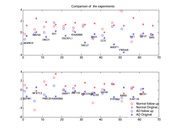
Figure 5.a : Comparison of the original and follow up experiment data.
Numeric output can be found in the file figure5.mat
|
Variable |
Description |
|
meanR{1} (matrix, cell array element) |
1977x1 normalized log-ratio average expression levels for Normal brain |
|
meanR{2} (matrix, cell array element) |
1977x1 normalized log-ratio average expression levels for AD brain |
|
diffs40 (vector) |
List of the 40 most differentially expressed genes |
|
followupN (31x1 vector) |
Vector of expression levels in the follow-up experiment in the Normal brain |
|
followupA (31x1 vector) |
Vector of expression levels in the follow-up experiment in the AD brain |
|
C (scalar ) |
Correlation coefficient |
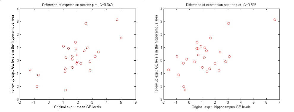
Scatter plots of the gene expression differences.
Left plot is the mean expression in the original experiment vs.
hippocampus in the follow up experiment. C = 0.65
Right plot is the hippocampal expression in the original experiment vs.
hippocampus in the follow up experiment. C = 0.59
This Article
-
doi: 10.1101/gr.204102 Genome Res. February 1, 2002 vol. 12 no. 2 244-254

