
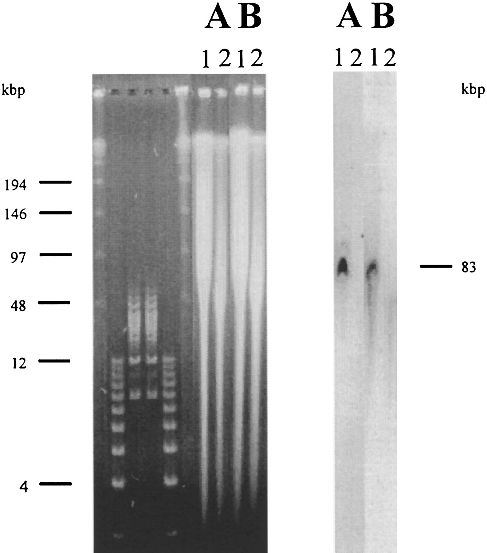
Direct measurement of gap 2 with genomic Southern blot hybridization. (Left) Photograph of a pulsed-field gel stained with ethidium bromide. Unlabeled lanes contain markers with some band sizes (in kb) are indicated. The next four lanes contain genomic DNA prepared in agarose blocks and digested with XhoI. (Lane 1) WA 17 (hybrid line containing only human chromosome 21 on mouse A9 background); (lane 2) A9 control. A and B are identical pairs of lanes run on the same gel, blotted, separated by cutting of the blotted membrane, and hybridized to end probes from B39I12 (SP6 end) and 190N10 (T7 end), respectively. (Right) Printout of a PhosphorImager scan of the hybridized membranes (B39I12, A;190N10 B). Both ends were generated with a modified vectorette–PCR technique (see Methods); B39I12 SP6 end was a 750-bp fragment amplified from the vectorette library generated withPstI; the 190N10 T7 end was a 420-bp fragment amplified from the vectorette library generated with HindIII.


