
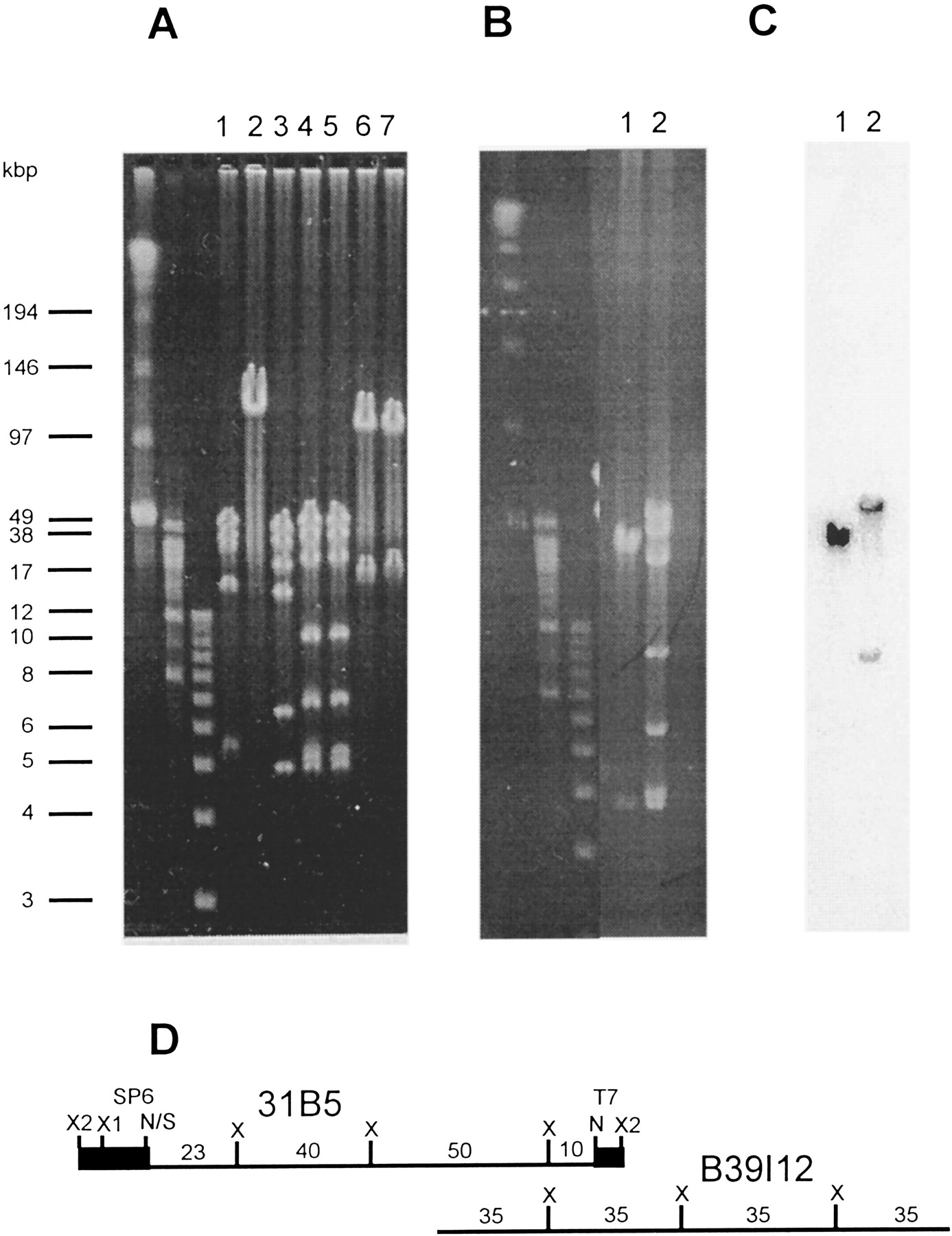
Restriction mapping and verification of the degree of overlap. (A) Restriction mapping of a PAC. The PFGE of the PAC clone 31B5. (Lanes 1) XhoI; (lane 2)SalI; (lane 3) XhoI–SalI; (lane4) NotI–XhoI–SalI; (lane5) NotI–XhoI; (lane 6)NotI; (lane 7) NotI–SalI. (B) PFGE of two overlapping clones from the MTP. (Lane 1) BAC B39I12 cut with NotI–XhoI–SalI; (lane 2) PAC 31B5 (restriction mapped in A) cut withNotI/XhoI. The first three un-numbered lanes inA and B contain molecular weight markers (see Methods), whose fragment sizes in kb are indicated on theleft. (C) Verification and sizing of the overlapping segment; the Southern blot of B hybridized to the insert from the BAC B39I12. (D) Restriction map of PAC 31B5 and the overlap of the two clones as deduced from A, B, andC. (N) NotI; (S) SalI; (X1 and X2) twoXhoI sites in the vector. SP6 and T7 are the RNA polymerase promoters flanking the BamHI cloning site of the PAC vector.


