
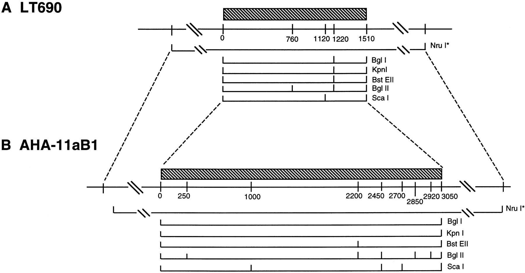
Restriction maps of centromeric α-satellite arrays and adjacent DNA in LT690 (A) and AHA–llaB1 (B). Distances are shown in kilobases; α-satellite sequences are present on those fragments indicated above by hatching. Rare cutting restriction enzymes such asNruI (*) have sites outside the array edges; these enzymes yield restriction fragments containing the entire α-satellite array along with variable amounts of flanking sequence. Fragment sizes obtained by digestion with these restriction enzymes are listed in Table 1, although the positions of these sites relative to one another was not determined. The broken lines point out the similar features of the DNA near the two chromosome centromeres: the clustering of common cutting restriction fragments at the array edges, and the positions of rare cutting restriction sites relative to these edges.


