
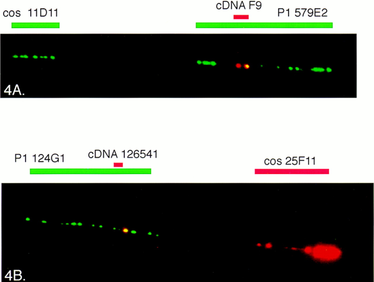
Figure 4.
The fiber FISH image of the region between markers D21S25and D21S154 (A) and around markerD21S171 (B). (A) A biotin-labeled candidate cDNA F9 (red) is visualized by tyramide detection on P1 clone 579E2 (long green signal), and a digoxigenin-labeled cosmid (cos) 11D11 (green) shows the orientation of the clones (centromere–cos 11D11–P1 579E2/cDNA F9–telomere). (B) A biotin-labeled IMAGE clone 126541 (red) is positioned on P1 124G1 (green) and cosmid 25F11 is visualized by tyramide-based detection to confirm the orientation of the clones (centromere–P1 124G1–cDNA 126541–cos 25F11–telomere).


