
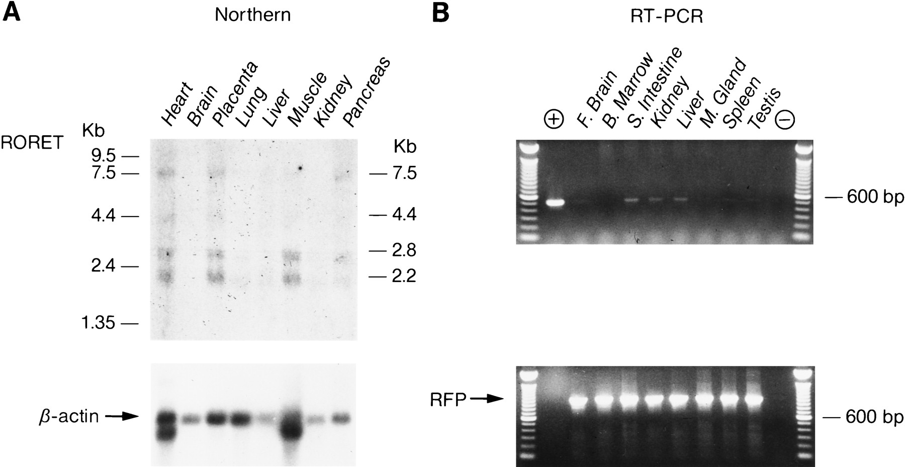
Expression analysis of the RoRet gene. (A) Northern blot analysis of the RoRet gene. The RoRet cDNA hybridizes to four different transcripts, ranging from 7.5 to 2.2 kb. Autoradiography was performed for 4 days. The rehybridization of the blot with a β-actin probe shows the variation in poly(A)+ RNA between the lanes. Autoradiography was for 1 hr. (B) RT–PCR analysis of the RoRet gene. Included in the (+) lane is a RoRet cDNA positive control. Weak amplification of the correct size is observed in the small intestine, kidney, and liver. The other tissues are negative as is the no-DNA control lane (−). The RFP primers show the integrity of the DNA.


