
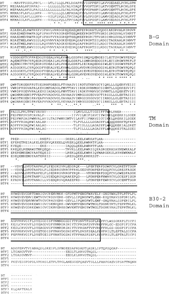
Alignment of the predicted amino acid sequence of the BTF proteins. Sequences were aligned in a pair-wise fashion using CLUSTAL to deduce the most parsimonious arrangement and are presented as such. The “stars” under the alignment represent those amino acids conserved in all six proteins, the “dots” represent conservative amino acid substitutions. Boxed are the regions within the proteins that correspond to three conserved motifs: (1) the B-G domain, (2) the transmembrane (TM) domain, and (3) the B30-2 exon domain. The sequences for all BTF cDNAs have been deposited in GenBank (accession nos: BTF1,U90543; BTF2, U90550; BTF3, U90548; BTF4, U90546; and BTF5, U90552).


