
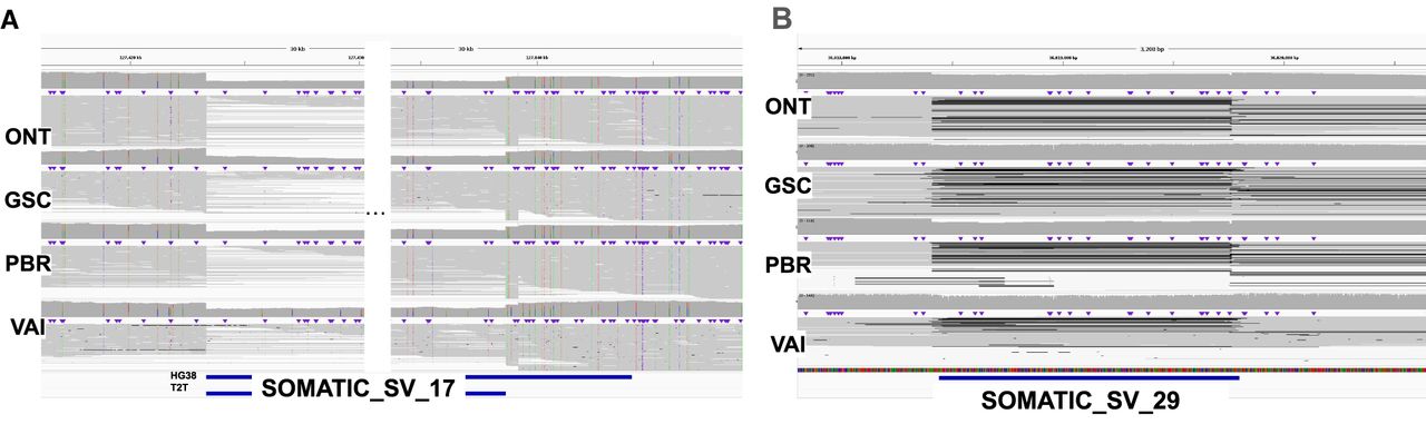
Figure 2.
Differences between the benchmarks based on the used reference. (A) IGV screenshot of an SV located in Chr7 that was reported with different sizes in CHM13-T2T and GRCh38. (B) IGV screenshot of an SV located in Chr17 cataloged as FP according to the benchmark by Valle-Inclan et al., which we are able to detect in all four cancer samples.


