
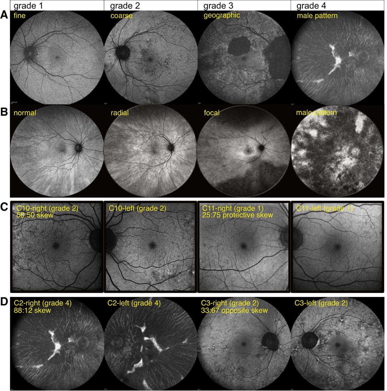
Clinical phenotypes of CHM and RPGR variant carriers and correlation with X inactivation skew. (A) Classification of retinal disease in female carriers of choroideremia based on 55° retinal fundus autofluorescence images, illustrative of the classification by Edwards et al. (2015): fine, coarse, geographic, and male-pattern degeneration. (B) Classification of retinal disease in female carriers of RPGR-associated X-linked retinitis pigmentosa based on 55° retinal fundus autofluorescence images, illustrating the classification by Nanda et al. (2018): normal, radial pattern, focal pigmentary retinopathy, and male-pattern degeneration. (C) Discordant clinical phenotypes of 34-year-old female C10 and her 59-year-old mother, C11, carrying the same pathogenic CHM variant. C10 presents with coarse (grade 2) phenotype, and C11 presents with fine (grade 1) phenotype. C11 had a pronounced buccal swab X inactivation skew (25:75) in favor of expressing the healthy allele, consistent with a milder disease phenotype. The 30° fundus autofluorescence images of right and left eyes are depicted for both carriers. (D) Discordant clinical phenotypes of 73-year-old female C2 and her 64-year-old sister, C3, carrying the same CHM variant. C2 presents with male-pattern degeneration (grade 4; picture also used to illustrate the grade 4 classification in panel A), and C3 presents with a coarse (grade 2) phenotype. C2 and C3 had skews in opposite directions, but the direction relative to the variant could not be determined. The 55° fundus autofluorescence images of right and left eyes are depicted for both carriers.


