
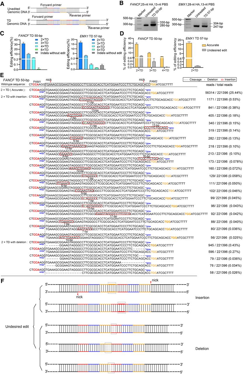
TD-PE generates a small fragment TD at the endogenous genomic locus. (A) PCR strategy for detecting the presence and frequencies of TDs. For each TD, a pair of primers flanking the duplicated fragment was designed. Successful TDs would generate bands with a larger size. (B) Agarose gel electrophoresis analysis of the presence of targeted TD on indicated loci. (C) HTS analysis of the frequencies of TD. The fragments containing TD in B were amplified by the HTS primers listed in Supplemental Table S5. Then the products were purified and subjected to HTS to analyze editing efficiency and product purity. Values and error bars reflect the mean ± SD of n = 3 independent biological replicates. (D) HTS analysis of the relative ratio of accurate and undesired editing outcomes of TD-PE. Values and error bars reflect the mean ± SD of n = 3 independent biological replicates. (E) HTS sequences showing the editing result of 2 × TD editing by TD-PE at the FANCF locus (reads with frequencies ≥0.02% were shown). The duplicated sequences are placed on separate lines and are indicated by blue curved arrows. Deletion, insertion, and base substitution are indicated by a black dash, a red box, or green letters, respectively. The insertion sequence of the red box was consistent with its 5′ upstream sequence. Note that sequences of ≥3 × TDs were displayed in Supplemental Figure S2. (F) Schematic diagram showing the pattern of insertion or deletion within undesired TDs, with the majority occurring at the boundaries of the duplications.


