
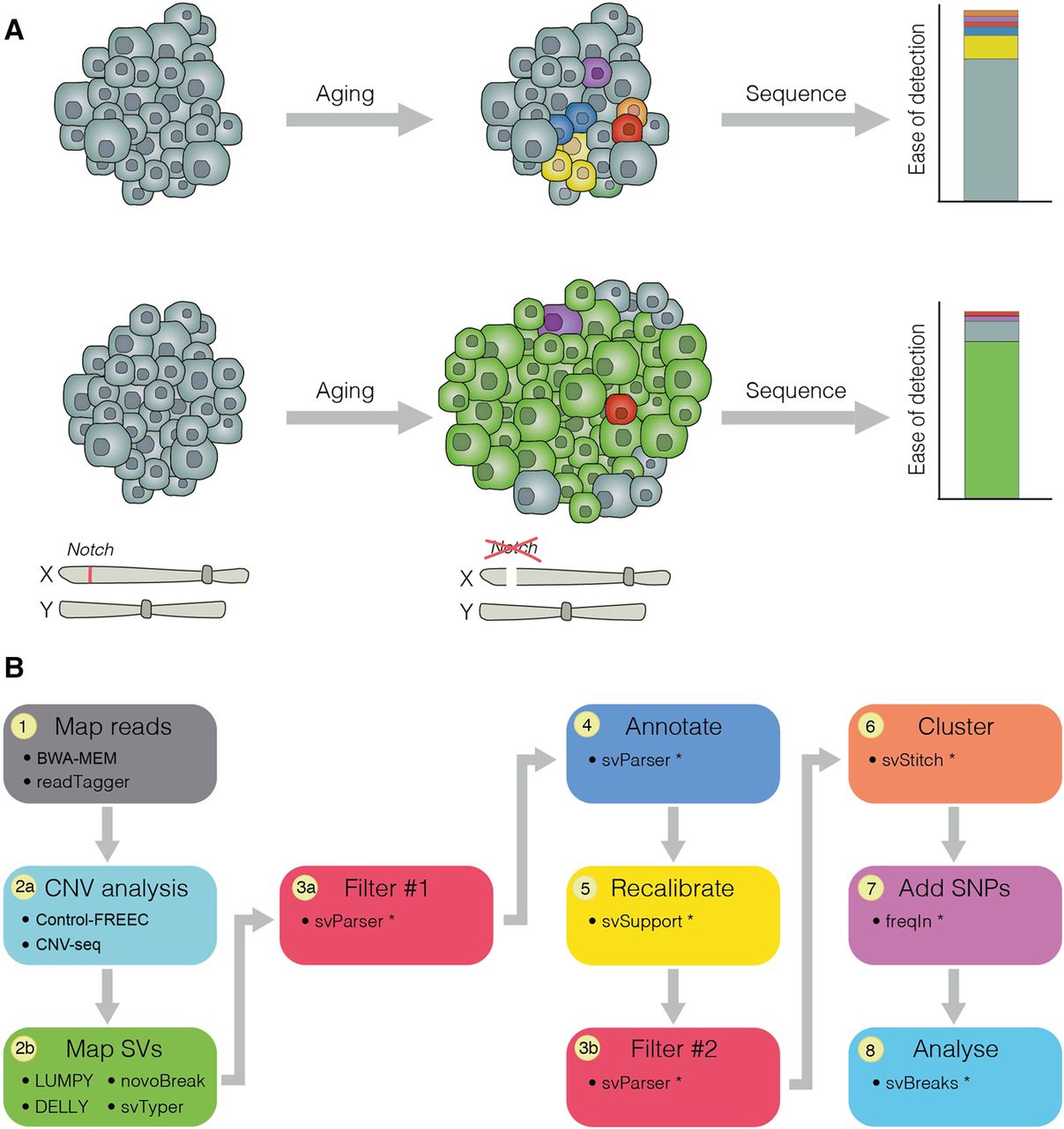
Clonal expansion of ISCs can be exploited to detect somatic mutations. (A) During aging, normal cells (gray) acquire somatic mutations (colored), typically restricted to small populations of cells. Bulk DNA-sequencing of such tissues fails to detect somatic mutations, as they are present in such small numbers. Somatic mutations occurring in an ISC (green) are inherited by the cell's progeny and, in the context of a neoplasm (e.g., as a result of loss of the X-linked tumor-suppressor gene Notch), are present in many cells within the tissue. As a result, sequencing of neoplasia increases the ability to detect somatic mutations in wild-type tissue. (B) A comprehensive bioinformatic pipeline was created in order to accurately detect and characterize structural variants from sequenced neoplasia. We have developed multiple packages to enable us to tag reads that map to multiple genomes (Siudeja et al. 2021), and filter and annotate structural variant breakpoints (svParser, svSupport, freqIn; Methods; Supplemental Methods). Our pipeline uses multiple approaches to detect structural variants and applies stringent filtering steps before annotating variants. Steps marked by an asterisk indicate bioinformatic tools developed for this study.


