
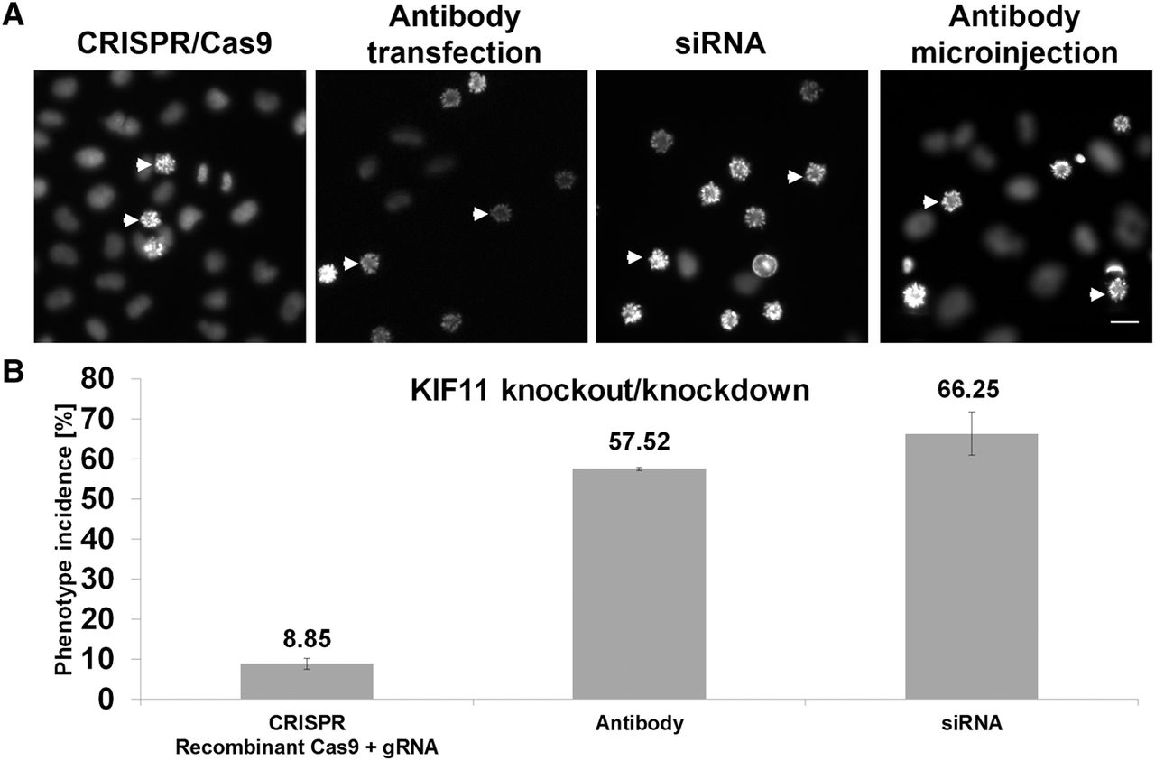
Comparison of KIF11 depletion by gene editing, RNAi, and antibody transfection. (A) Prometaphase arrest was obtained after solid-phase reverse transfection of HeLa cells: (left to right) with recombinant Cas9 + gRNA targeting KIF11, anti-KIF11 antibody, siRNA targeting KIF11, and anti-KIF11 antibody microinjection. Two example cells showing the prometaphase arrest phenotype are indicated by arrows in every image. (Scale bar) 20 µm. (B) Phenotype incidences after KIF11 knockout and knockdown. Bars represent average values derived from six independent replicas, and error bars represent the standard deviations. Phenotype analysis and quantification were performed as described in Methods. Transfection of the anti-KIF11 antibody was performed under the conditions described in Figure 1. The transfections were performed in 384-well plates and the microinjection was performed in a 35-mm petri dish.


