
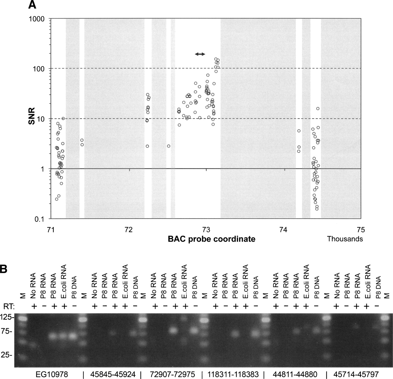
(A) Signal-to-noise ratio for human BAC P8 probes located on a 4-kbp stretch between coordinates 71 and 75 kbp. Transcription signals are detected throughout the BAC and are particularly strong in the 73-kb region. The double-sided arrow indicates the position of a -p RT-PCR product, amplified from an RNA preparation of DH10B transformed with human BAC P8. The gray areas are regions excluded from oligonucleotide design due to repeat content, self-folding, or elevated Tm. (B) Human BAC P8 regions exhibiting strong transcription signals were identified and used for RT-PCR primer design. This SYBR Green-stained agarose gel image shows corresponding RT-PCR amplicons from a DNase-treated RNA preparation from DH10B transformed with the whole human BAC P8.


