
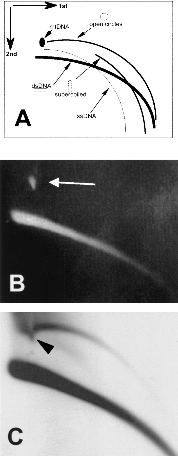
Two-dimensional gel analysis of extrachromosomal circular DNA in Drosophila embryos. (A) A schematic outline of the 2D gel electrophoretic patterns of genomic DNA generated by populations of linear and circular molecules (Cohen and Lavi 1996). Each arc consists of molecules sharing the same structure, but differing in mass. This analysis allows discrimination between double-stranded DNA (dsDNA), single-stranded DNA (ssDNA), relaxed (open) circular molecules, and supercoiled molecules. It also enables detection of the mitochondrial DNA (mtDNA). (B) Ethidium bromide staining of a 2D gel analysis of genomic DNA from early Drosophila embryos reveals an arc of the chromosomal linear DNA and a spot of the mtDNA (19.5-kb circular DNA, white arrow). (C) Hybridization with total genomic DNA probe reveals an arc of relaxed circles in addition to the massive arc of linear DNA. Note that the large quantity of mtDNA caused a local shift in the migration of the arc of the circular molecules (arrowhead), which extends beyond the mtDNA.


