
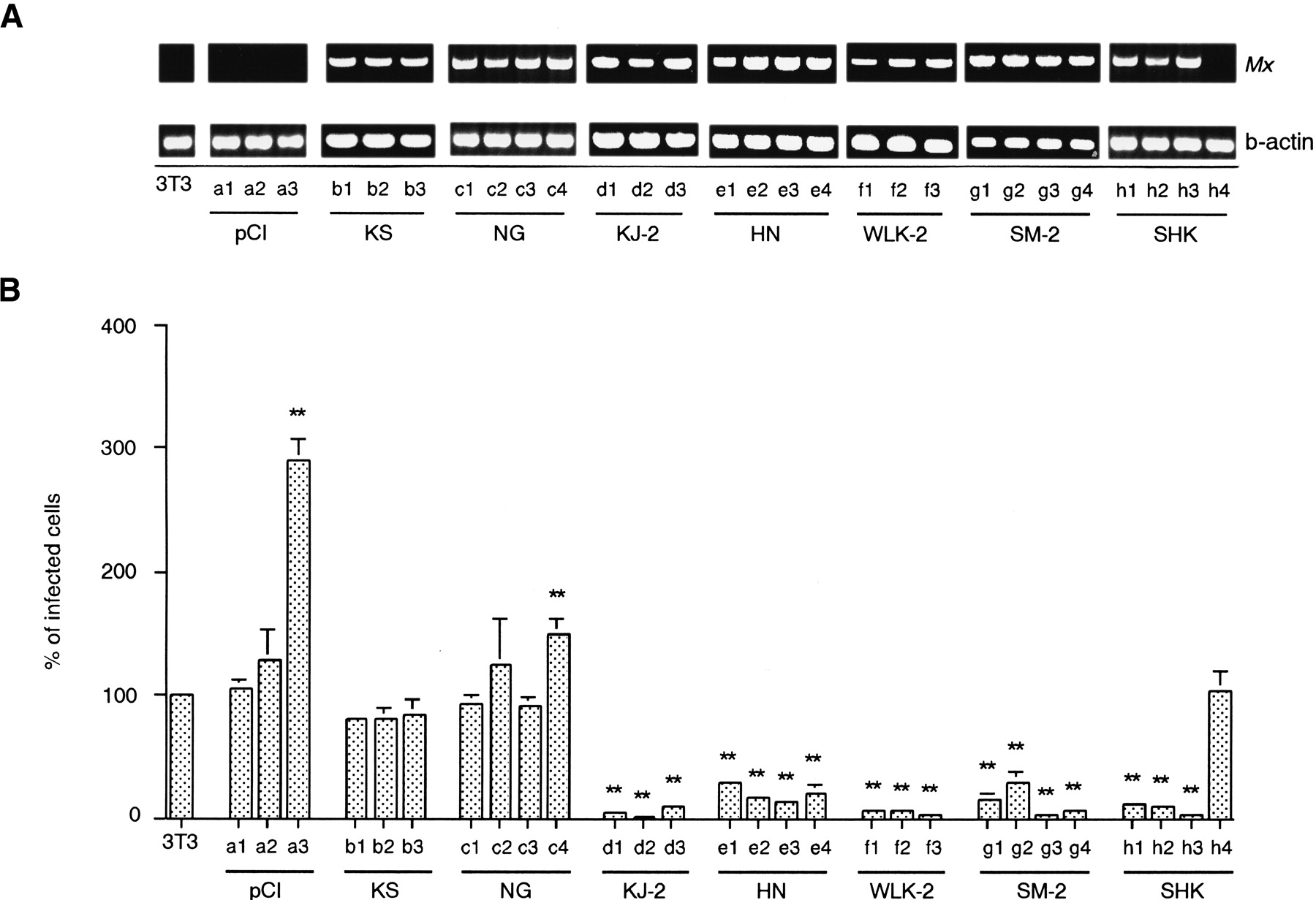
(A) The expression of Mx mRNA in each clone. Total RNA from transfected 3T3 cells was reverse transcribed and PCR amplified using Mx primers (top) and β-actin primers (bottom). 3T3, parental 3T3 cells; pCI (a1–3), control 3T3-pCI-neo; KS (b1–3), 3T3 cells expressing KS MxmRNA; NG (c1–4), 3T3 cells expressing NG Mx mRNA; KJ-2 (d1–3), 3T3 cells expressing KJ-2 Mx mRNA; HN (e1–4), 3T3 cells expressing HN Mx mRNA; WLK-2 (f1–3), 3T3 cells expressing WLK-2 Mx mRNA; SM-2 (g1–4), 3T3 cells expressing SM-2 Mx mRNA; and SHK (h1–3; h4 failed to express theMx mRNA), 3T3 cells expressing SHK Mx mRNA. An aliquot of each PCR product was electrophoresed on a 1.2% agarose gel and visualized with ethidium bromide. (B) The infectivity of VSVΔG*-G in Mx cDNA-transfected cell lines. The infectivity on parental 3T3 cells is expressed as 100%. Shown are mean value ± standard errors of the means (n = 10). Significance levels atP < .01 (**) compared with 3T3 cells are indicated.


