
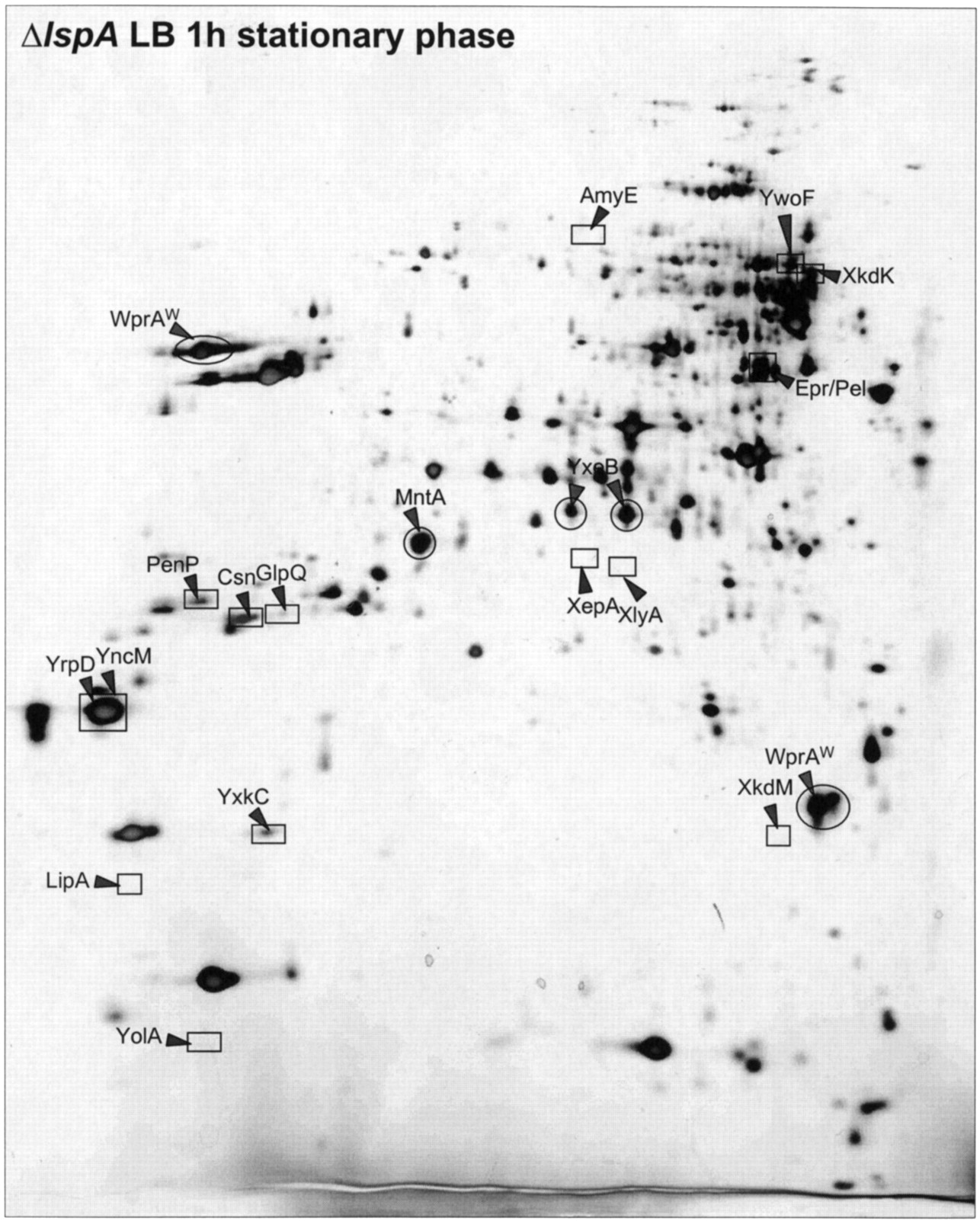
The extracellular proteome of SPase II mutant cells of B. subtilis. Cells of B. subtilis ΔlspA, which lack SPase II, were grown in L-broth and the proteins in the growth medium were harvested 1 h after entry into the stationary phase. After precipitation with TCA, the extracellular proteins were separated by 2D PAGE as described in the Methods section. The proteins identified by mass spectrometry are indicated. Extracellular proteins that are present at elevated levels are encircled, and extracellular proteins that are present at reduced levels are boxed (see also the listing in Table 3). This gel is available in the Sub2D proteome database (http://microbio2.biologie.uni-greifswald.de:8880/).


