
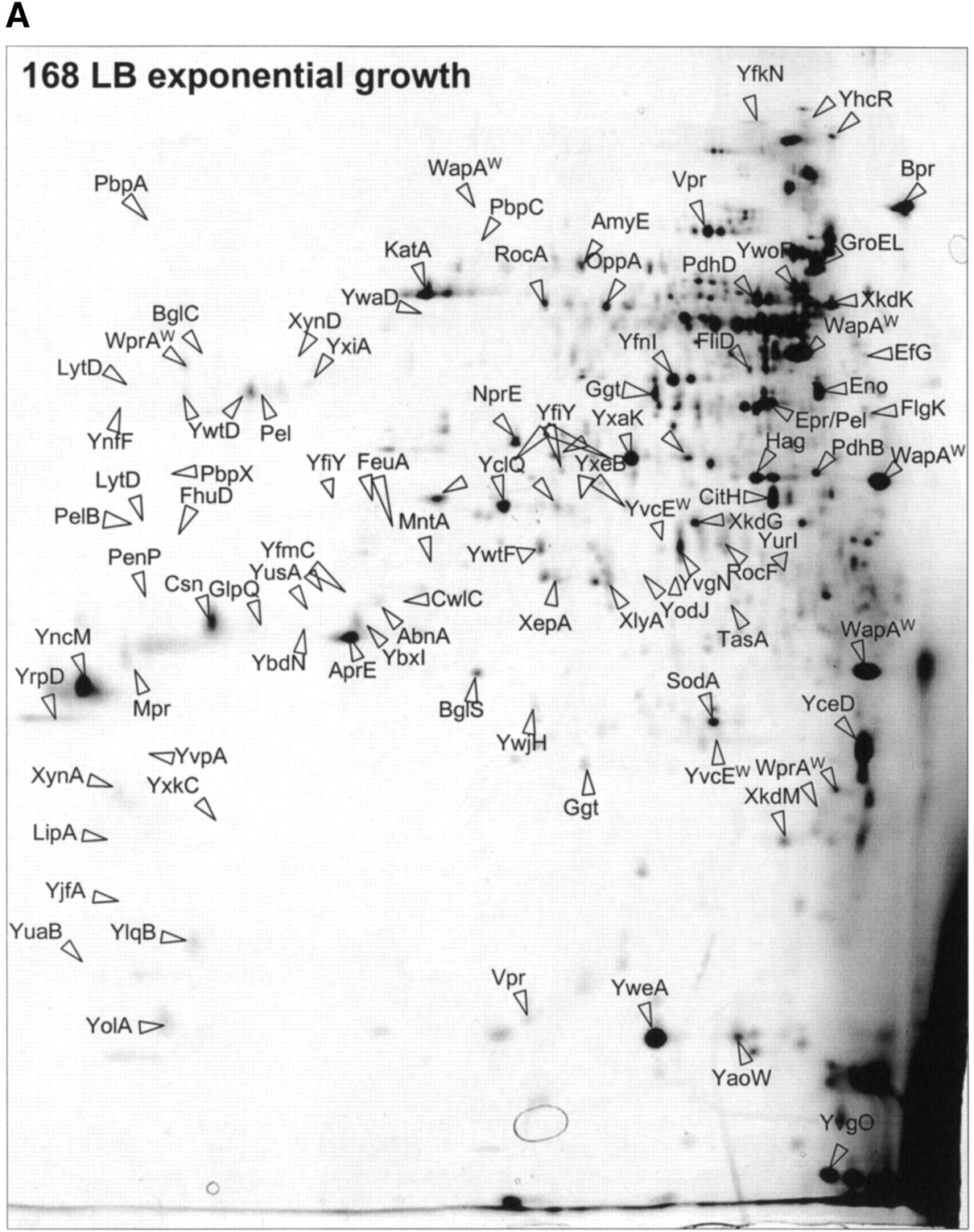
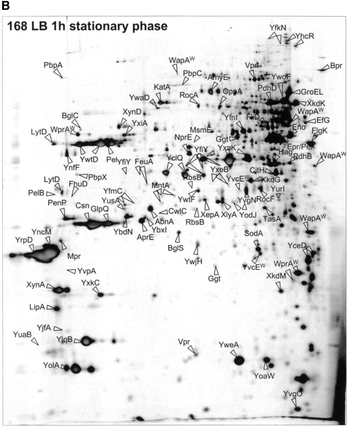
Figure 1.
The extracellular proteome of B. subtilis 168. Cells ofB. subtilis 168 were grown in L-broth and proteins in the growth medium were harvested during exponential growth (OD540 = 0.4) (A) and 1 h after entry into the stationary phase (B; master gel for the extracellular proteome). After precipitation with TCA, the extracellular proteins were separated by 2D PAGE as described in the Methods section. The proteins identified by mass spectrometry are indicated on the gel and listed in Table 1. These gels are available in the Sub2D proteome database (http://microbio2.biologie.uni-greifswald.de:8880/). (See following page for panel B.)


