
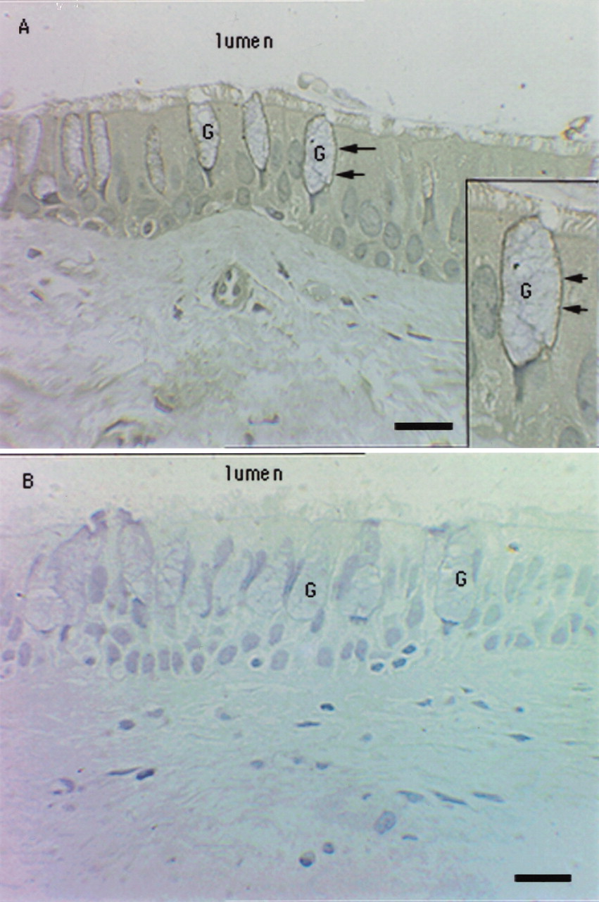
Figure 8.
Photomicrograph of T4 immunostaining in a representative section of a bronchial biopsy from an asthmatic subject (A). Also shown is photomicrograph of a representative section stained with negative control (MOPC-31c) (B). The figure shows distinct T4 staining along the basolateral membrane of goblet cells. Arrows point to basalateral immunostaining of goblet cells (G). No other epithelial cells stained positively. The inset in panel Ashows a magnified field of the same specimen. Bar = 20μM.


