
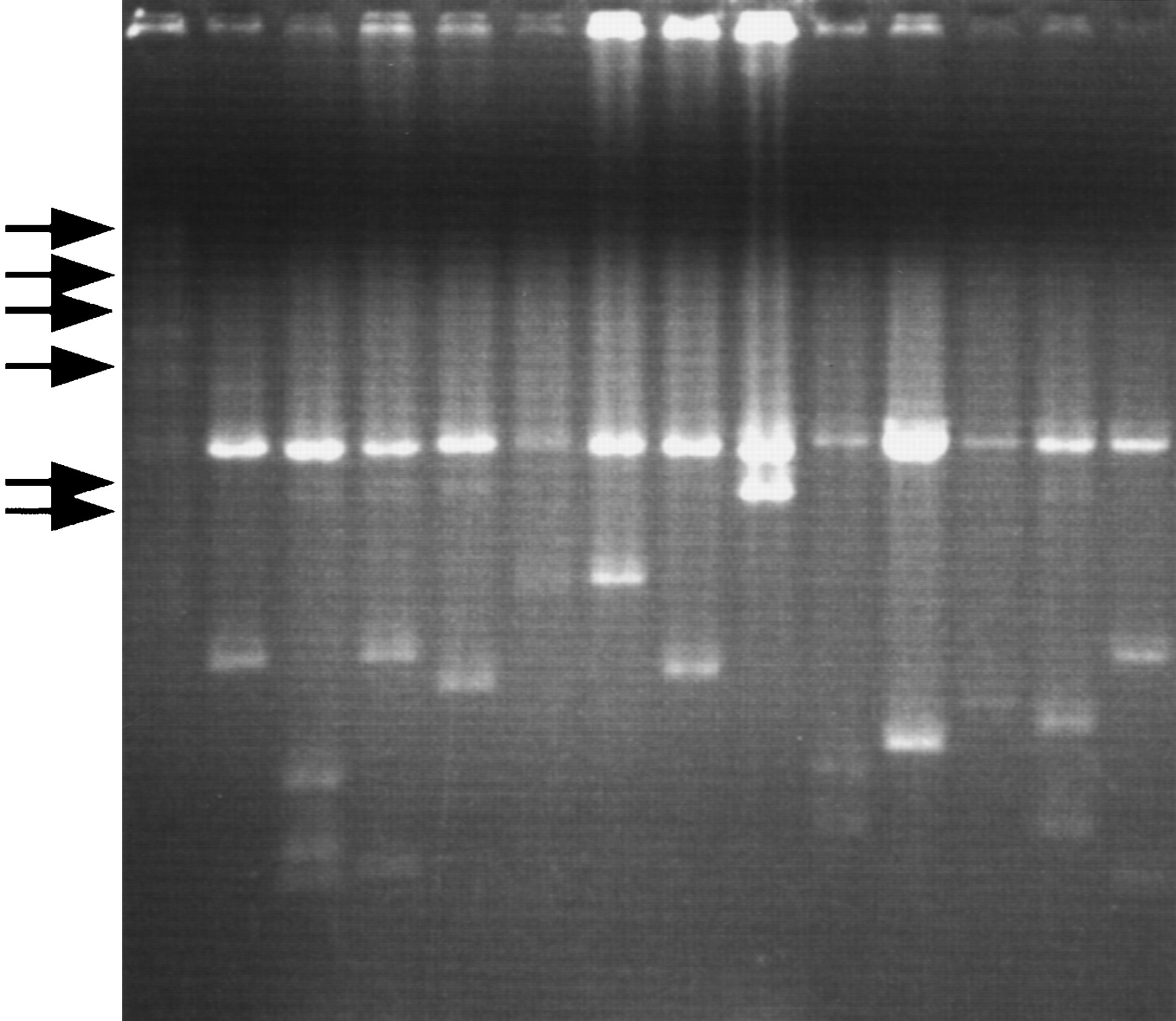
Figure 3.
Agarose gel electrophoretic patterns of plasmid DNAs prepared by this system. cDNAs were cloned into λ-ZAPII and excised in E. coli SOLR cells. The plasmids were digested with PvuII, and subjected to electrophoresis. (Arrows) Locations of DNA size marker (λ -HindIII digest).


