
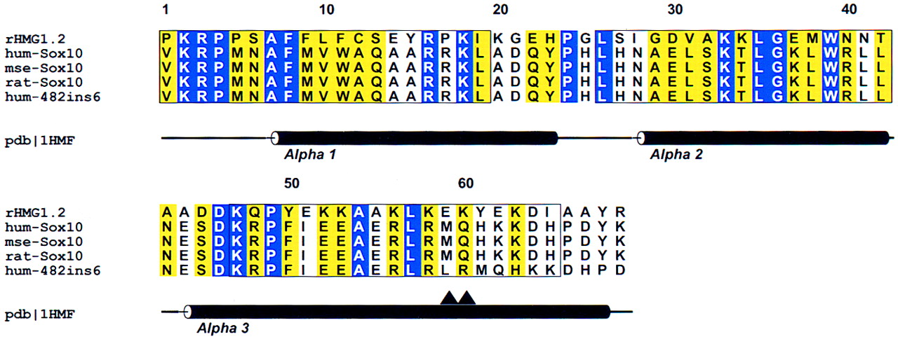
Structural alignment of the HMG-1 box domains of human, mouse, and rat Sox10 proteins. The sequence of HMG-1 box 2 from rat whose NMR structure was used as the basis for the threading experiments (Weir et al. 1993) is shown in the first line of the alignment (rHMG1.2). (Blue) Positions exhibiting absolute identity; (yellow) conserved positions. (▴) The positions of the 2-residue Leu–Arg insertion in human 482ins6. Positions of the secondary structural elements found in the HMG-1 box NMR study are shown below the alignment. Core segments defined for the threading algorithm (Bryant and Lawrence 1993) are boxed. ALSCRIPT V. 2.0 (Barton 1993) was used to format the alignment.


