
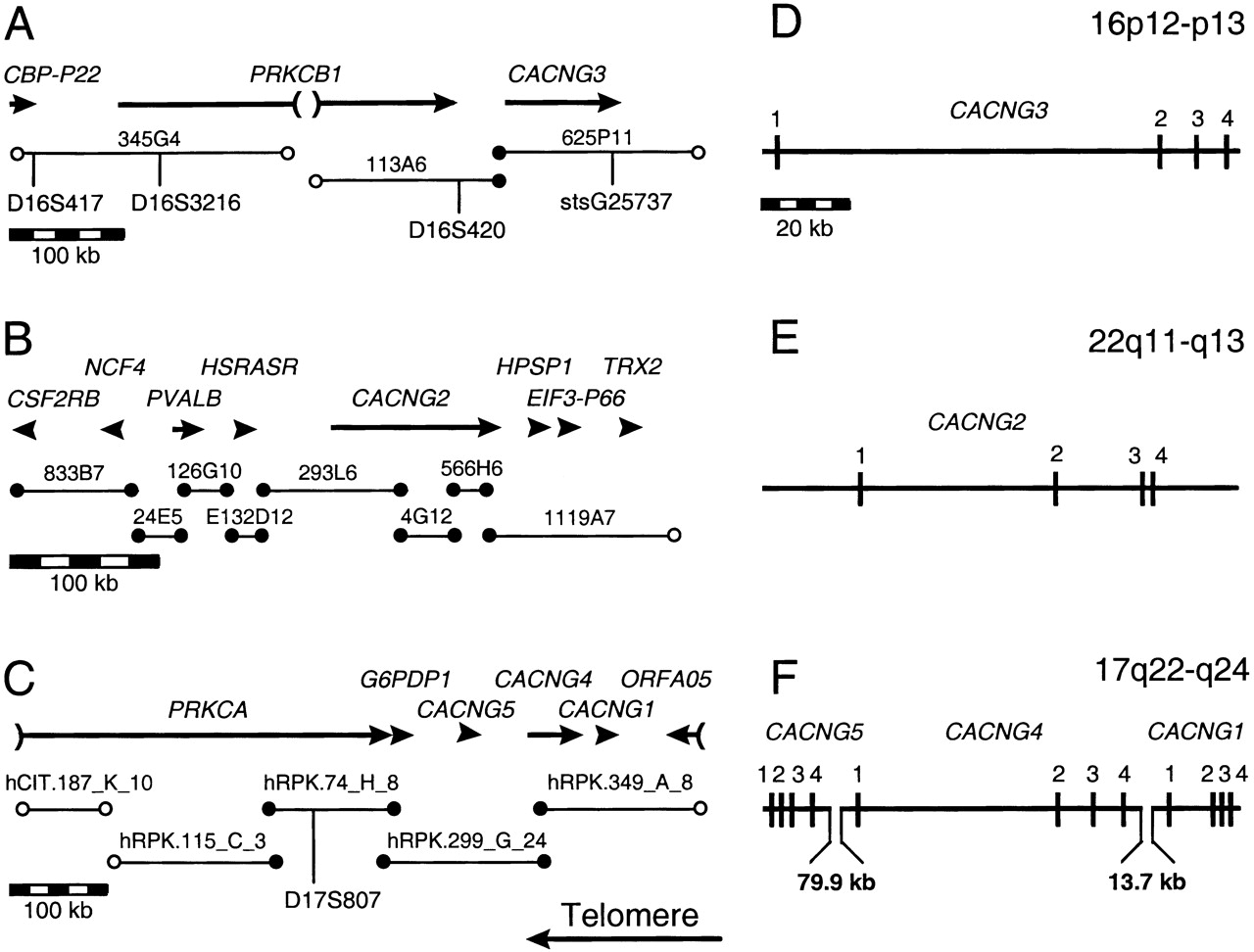
(A–C) Physical maps of the region surrounding the human Ca2+ channel γ subunit genes. Genomic sequences are indicated by thin lines below the name of the associated clone, with selected markers shown below. (●) Sequence overlap; (○) end sequences associated only with a single parent clone in GenBank. Characterized genes are represented by thick lines, with transcriptional orientation indicated by arrowheads. PRKCB1 is interrupted by a gap in the genomic sequence of unknown length. Only the 3′ ends of PRKCA and ORFA05 are currently represented in GenBank. (CBP–P22) calcineurin-related gene; (PRKCB1) protein kinase C β1; (CACNG3) calcium channel γ3; CSF2RB colony-stimulating factor 2 receptor β; (NCF4) neutrophil cytosolic factor 4; (PVALB) parvalbumin; (HSRASR) similar to H. sapiens RAY1gene; (CACNG2) calcium channel γ2; (HPSP1) Hermansky–Pudlak syndrome pseudogene; (EIF3–P66) eukaryotic translation initiation factor 3, subunit 7 (ζ, 66/67 kD); (TRX2) thioredoxin 2; (PRKCA) protein kinase C α; (G6PDP1) formerly G6PDL, glucose-6-phosphate dehydrogenase pseudogene 1; (CACNG5) calcium channel γ5; (CACNG4) calcium channel γ4; (CACNG1), calcium channel γ1; (ORFA05) hypothetical myeloid cell line protein 5. StsG25737 represents an EST of CACNG3. Complete clone names and database accession numbers are given in Methods. (D–F) Comparison of the intron–exon structure ofCACNG1–5. The scale bar in D applies toD–F.


