
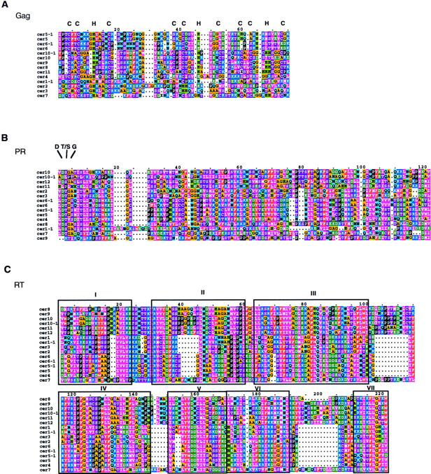
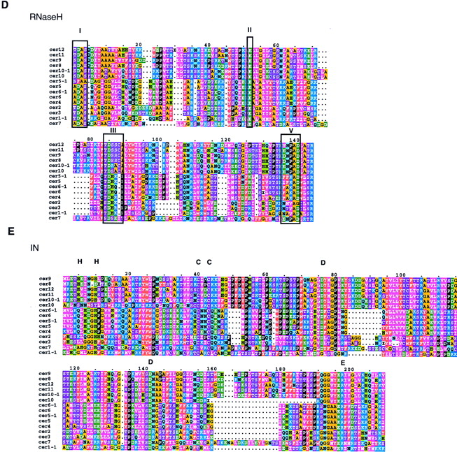
Cer protein alignments. The RT (C) of eachCer was identified according to the motifs previously designated by Xiong and Eickbush (1990). The additional protein domains (A = Gag, B = PR, D = Rnase H, andE = IN) were identified in a like manner using domains previously established by McClure (1991). The sequences were aligned using CLUSTAL X as described in Methods. Residue coloring was performed by MacBoxshade v2.01 and is based on the similarity scheme (F,W,Y), (I,L,M,V), (P), (D,E), (G,A), (S,T,C), (N,H),(R,K), (Q). Members of a similar residue group are the same color. In-frame termination codons or frameshifts are depicted by an X.


