
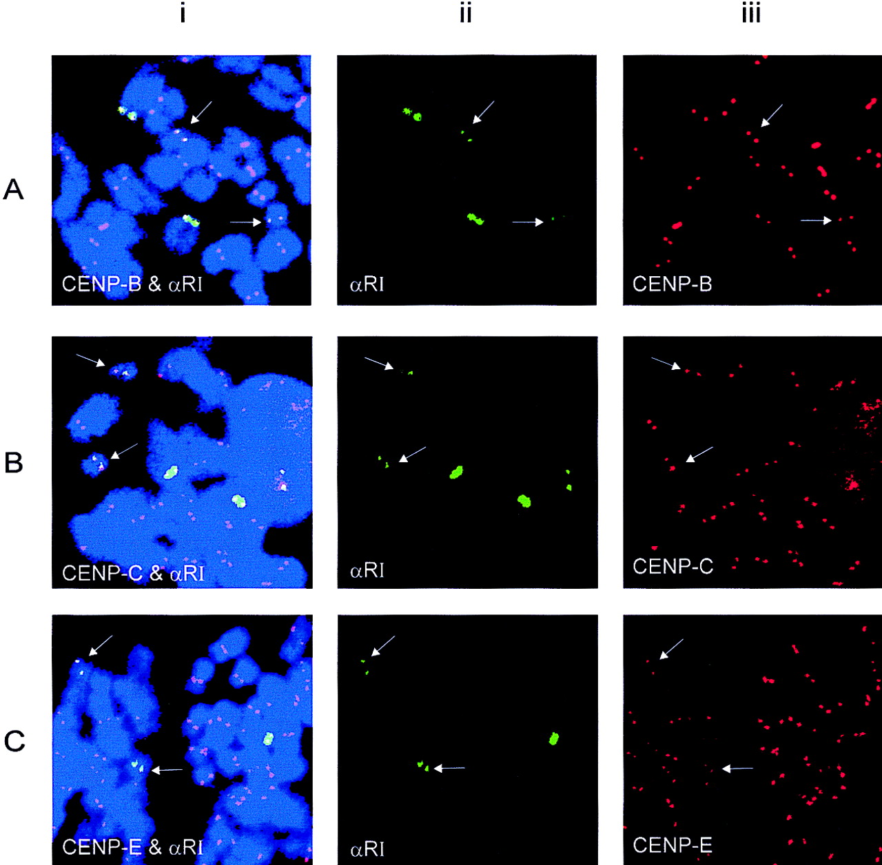
Figure 4.
Combined immunocytochemical and FISH analyses for the detection of CENP-B (A), CENP-C (B), and CENP-E (C) with the αRI probe. The cell line contains two low-αRI chromosomes 21 (arrows). (i) Combined images of centromeric protein signals (red) and the αRI signals (green); (ii) split images of i showing the FITC signals for the αRI domain; (iii) split images of i showing the Texas Red signals for the centromeric proteins. Nuclei and chromosomes are counterstained with DAPI and pseudocolored blue.


