
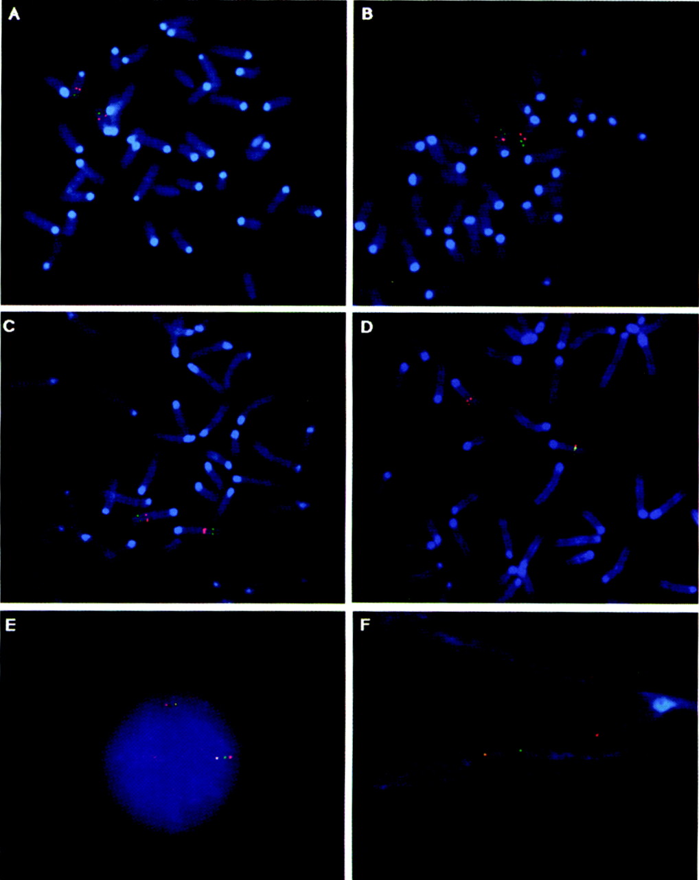
Figure 1.
FISH on normal mouse metaphase chromosomes, interphase nuclei, and extended chromatin. Brca1 (FITC, green fluorescence) was used as a marker for chromosome 11, and colocalized on metaphase spreads to chromosome 11 with: (A) Lis1; (B)Mnt/Rox; (C) 14-3-4ε; (D)Lis1, Mnt/Rox, and 14-3-3ε (rhodamine, red fluorescence). (E) Gene order was established by three–color FISH on mouse interphase nuclei: Lis1 (red); Mnt/Rox(green); 14-3-3ε (orange, mix of biotin- and digoxigenin-labeled DNA, 1:1 ratio). (F) FiberFISH was used for evaluation of relative distances between Lis1(orange), Mnt/Rox (green), and 14-3-3ε (red).


