
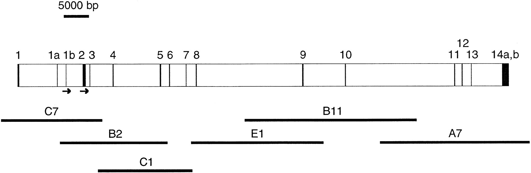
Figure 1.
Genomic structure of the EXT2 gene. The 16 exons are represented by solid boxes. Exons 1a and 1b are alternatively spliced exons. The 3′ UTRs 14a and 14b represent alternative polyadenylation sites (see text for details). The initiation codons are indicated by arrows. The cosmids used for DNA sequencing are shown below the map. Their placements reflect their exon content, but they are not necessarily to scale.


