
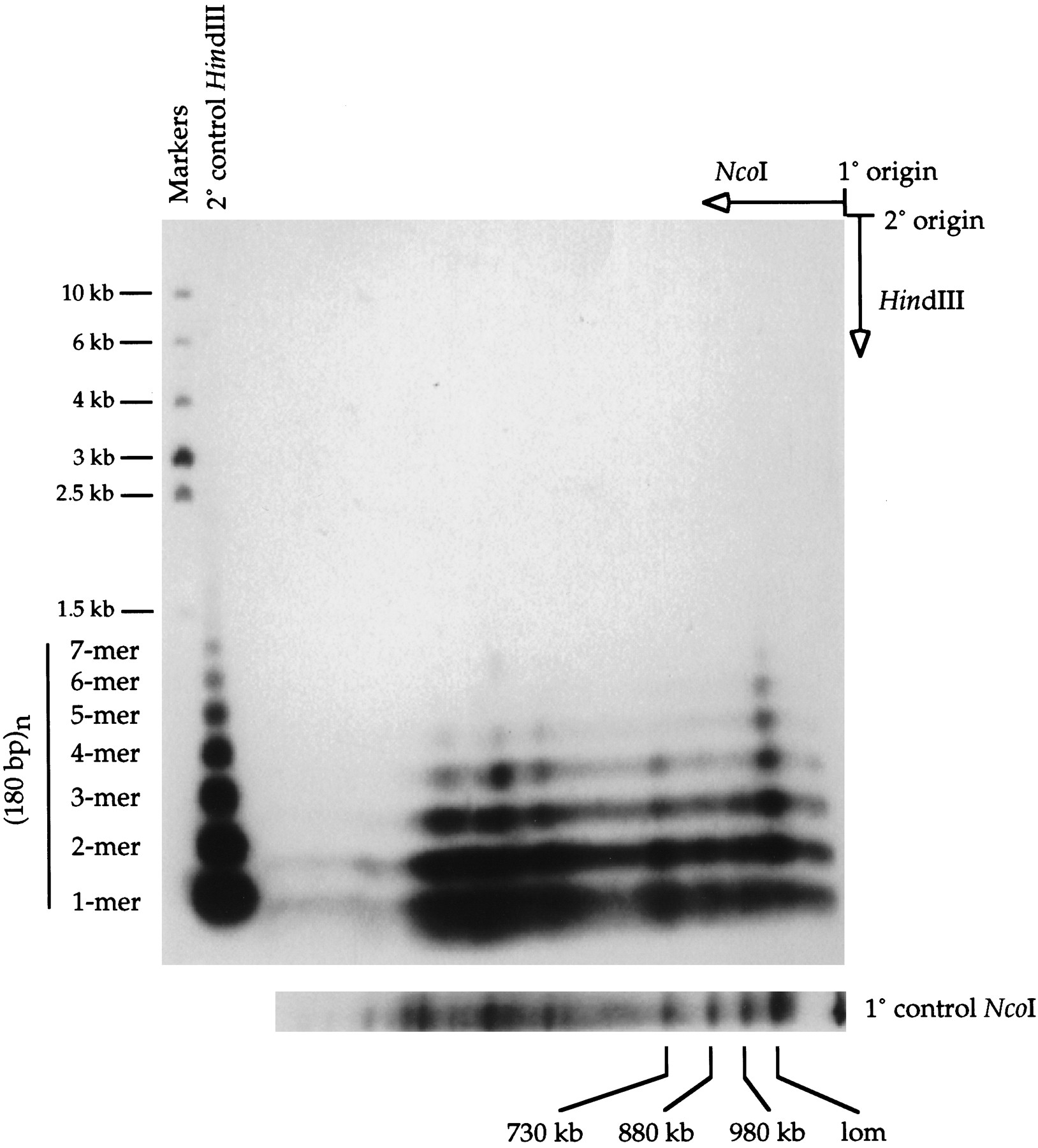
Two-dimensional gel analysis of large NcoI fragments containing 180-bp centromere repeats. A high-molecular-weight genomic DNA sample from A. thaliana strain Landsberg erecta was digested with NcoI and size-fractionated by pulsed-field gel electrophoresis in the first dimension. An excised gel slice containing the separated NcoI fragments was then incubated withHindIII and placed horizontally at the origin of a 1% agarose gel. After conventional electrophoresis in the second dimension, the fragments were transferred to a nylon membrane and detected by hybridization with a radiolabeled 180-bp repeat probe.HindIII-digested Landsberg erecta genomic DNA was run in the second dimension as a control (shown at left with molecular weight markers). Repeat multimers up to 7-mers were detected. The origin of the second-dimension HindIII control lane was offset slightly from the first-dimension gel slice, accounting for the faster apparent migration of the sample vs. control multimers. A separate first-dimension NcoI control lane is shown at thebottom of the figure. (lom) Limit of mobility.


