
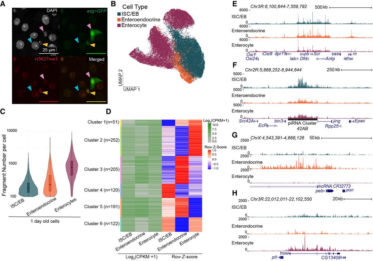
Repressed landscapes for cell types in the Drosophila gut. (A) Immunofluorescence image of the gut in a young esgGal4/CyO; UAS-GFP/TM6B female. GFP (green) marks stem cells and enteroblasts; red anti-H3K27me3, DAPI in gray. Pink arrowheads point to ISC/EBs; blue arrowheads point to enterocytes; and yellow arrowheads point to enteroendocrine cells. Enterocytes are the large DAPI-stained nuclei (blue arrow). Enteroendocrine cells are identified by their small nucleus size in relation to the larger enterocytes and low GFP signal (yellow arrows). (B) UMAP of H3K27me3 signal in gut cells from 1-, 15-, and 40-day-old females. Clusters are assigned cell types by a low chromatin silencing score (CSS) at marker genes. (C) Violin plots of the distribution of fragments per cell for 1-day-old cell types. (D) Log2-transformed counts per kilobase per million (CPKM)–normalized and Z-score-normalized heatmaps from differential analysis of H3K27me3 over genes between 1-day-old cell types. (E–H) UCSC Genome Browser tracks of repressed domains in the three cell types in young guts. (E) The ANTP-C domain is shared between all three cell types. (F–H) A domain encompassing the piRNA cluster 42AB is present only in ISC/EBs (F), a domain encompassing the lncRNA:CR32773 gene is present only in enteroendocrine cells (G), and a domain encompassing the how gene is present only in enterocytes (H).


