
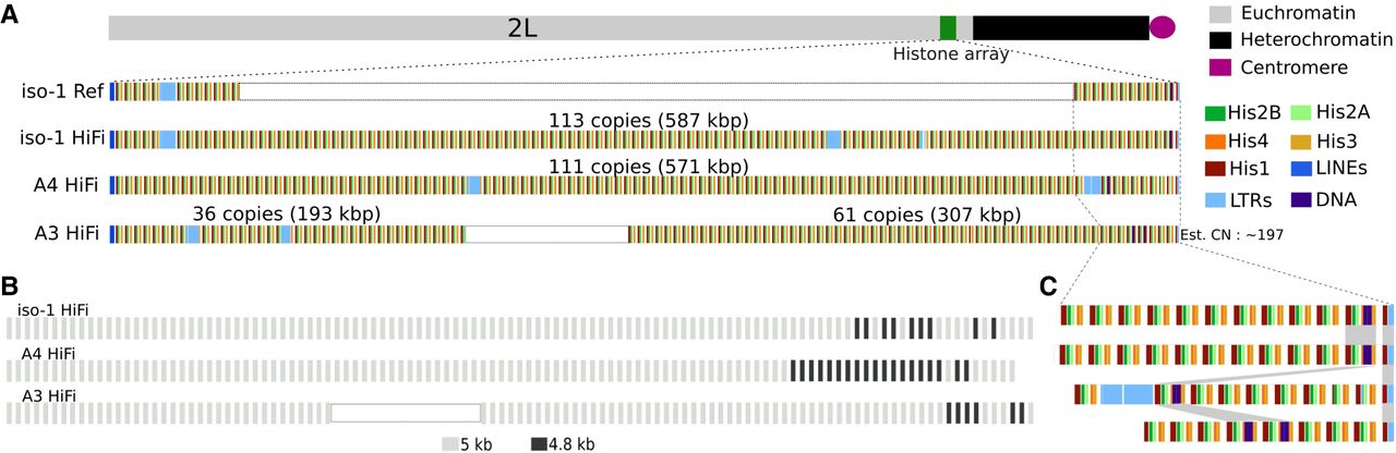
Maps of the newly assembled histone locus in Drosophila melanogaster. (A) Overview of the structure of the histone locus. Top: Schematic illustration of the location of the histone locus relative to the rest of Chromosome 2L. Bottom: Map of location of elements in the histone cluster, including the five individual histone genes and various transposable elements. “iso-1 Ref” refers to the Release 6 community assembly and “iso-1 HiFi” indicates the assembly presented here. White rectangles for iso-1 Ref and A3 HiFi indicate gaps resized to facilitate comparison with the other two assemblies. (B) Location distribution of the major histone unit types (i.e., 5-kb and 4.8-kb). (C) Expanded map of the proximal end of the array. Order of strains follows panel A.


