
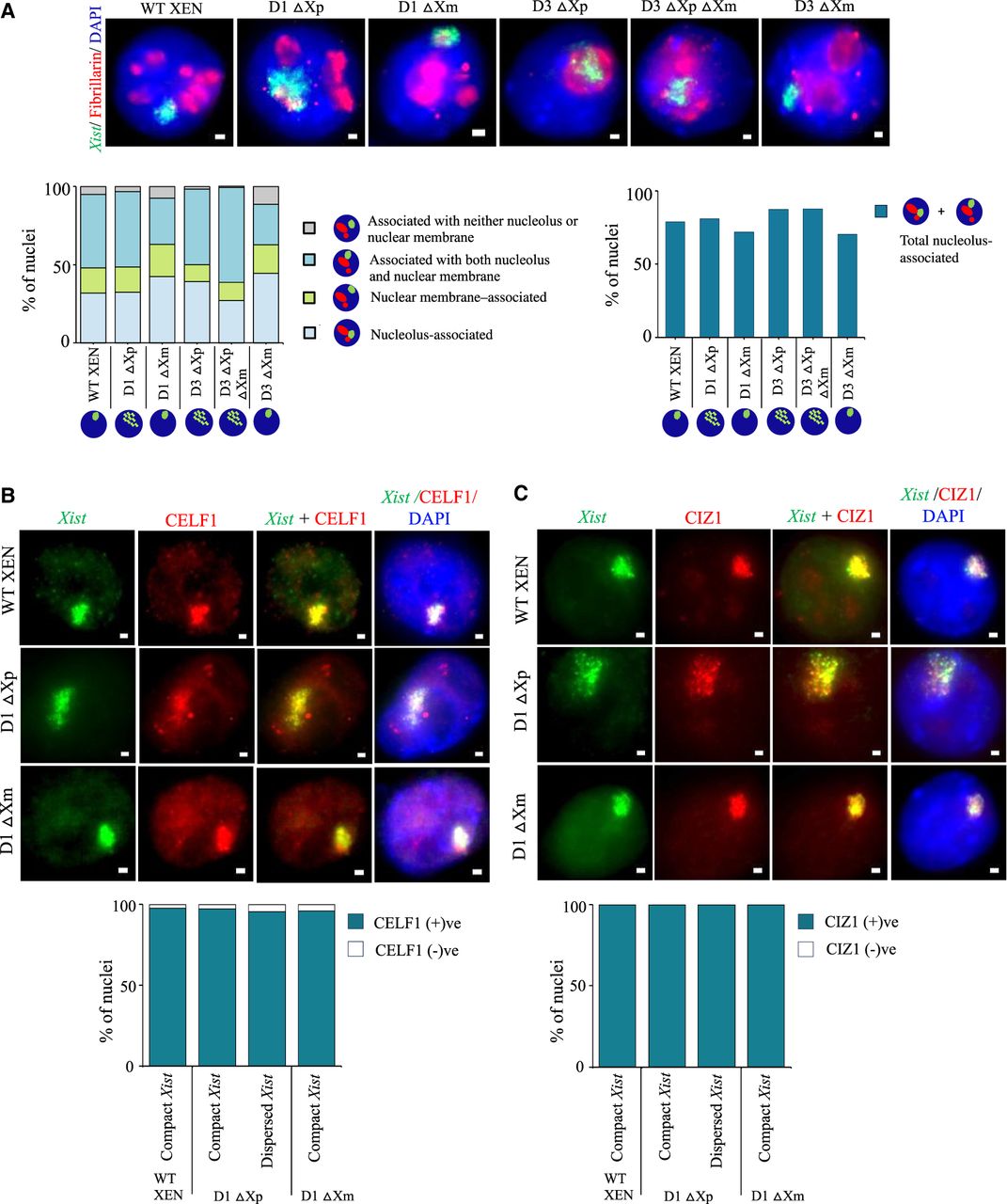
Xist-dispersed nuclei retains association with the nucleolus and do not show alteration in CELF1 and CIZ1 enrichment. (A) Top: Representative images of IF-RNA FISH for fibrillarin (red) and Xist (green) in WT, D1, and D3 deleted lines. Bottom:Quantification of the association between Xist cloud and fibrillarin signals in nuclei with compact and dispersed Xist clouds in the D1 and D3 deleted lines. n = 119, 157, 146, 120, 122, 148 (for each data point from left to right). Quantifications were performed based on single Z-planes. (B) Representative images for IF-RNA- FISH for CELF1(red) and Xist (green) in WT and D1 deleted lines. Bottom: Quantification of enrichment of CELF1 on the inactive X in compact versus dispersed Xist nuclei. n = 246, 110, 96, 101 (for each data point from left to right). (C) Images representing IF-RNA-FISH for CIZ1 (red) and Xist (green) in WT and D1 deleted lines. Bottom: Quantification of enrichment of CIZ1 on the inactive X in compact versus dispersed Xist nuclei. n = 128, 111, 120, 114 (for each data point from left to right). A single-stranded RNA probe was used to detect Xist.


