
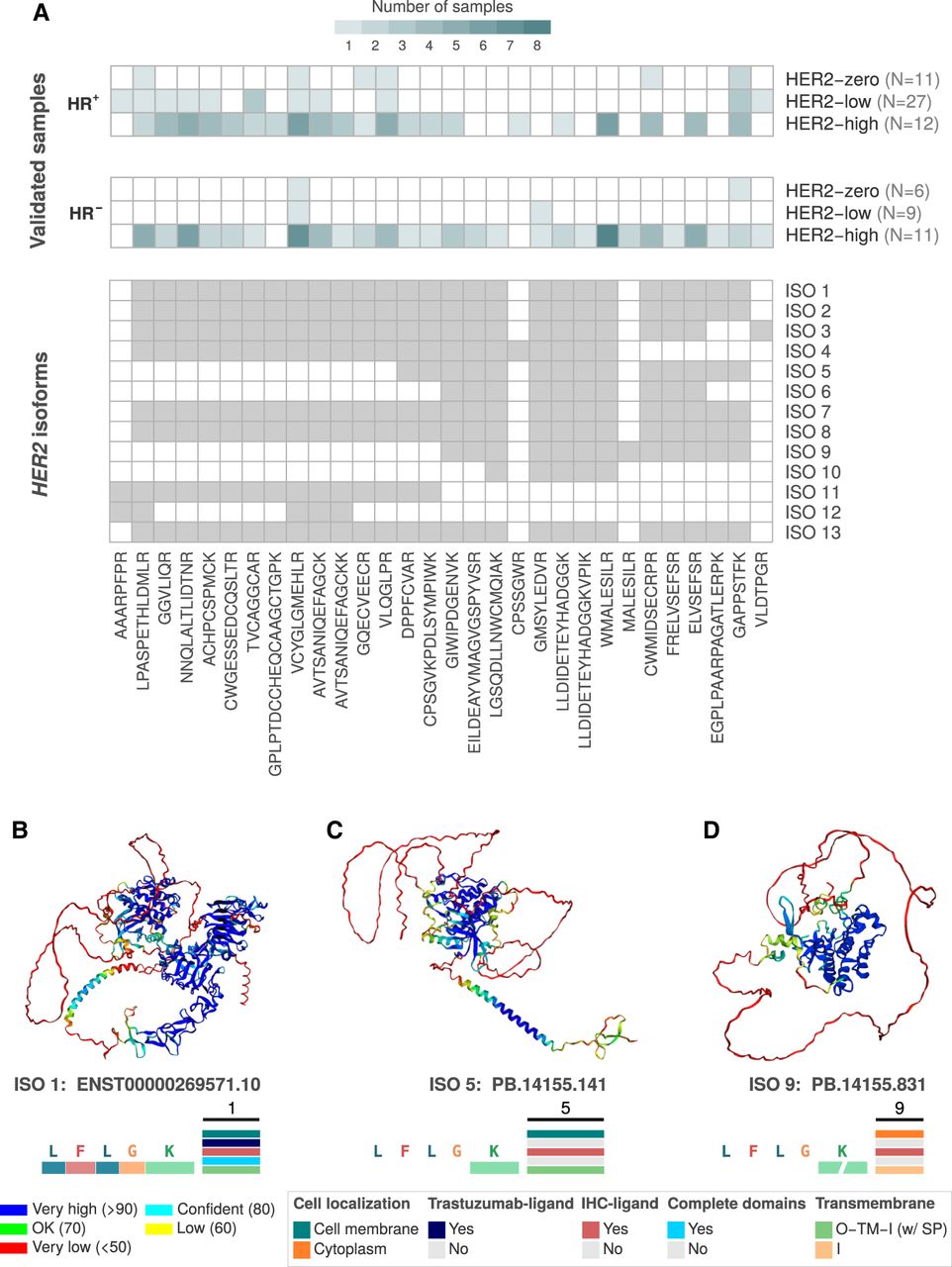
Mass spectrometry (MS) validation of HER2 isoform–derived proteins and their predicted 3D structures. (A) The top panels display the number of samples with MS confirmation of HER2 proteins, stratified by HER2 status (HER2-high, HER2-low, HER2-zero) and hormone receptor status (HR+ and HR−). Isoform-derived proteins are grouped from 1 to 13, and the validated peptides for each group are indicated in the lower panel. (B–D) AlphaFold2-predicted protein structures for HER2 isoforms. (B) The canonical isoform (ISO 1: ENST00000269571) with well-defined domain regions. (C) ISO 5 (PB.14155.141), a variant from group 5 with specific domain alterations affecting its cellular localization and trastuzumab-binding potential. (D) ISO 9 (PB.14155.831), an isoform with unique structural characteristics lacking complete domains, potentially affecting functional properties. Color codes in each structure represent pLDDT confidence scores for structural predictions and the corresponding HER2 protein domains, as the bottom legend indicates. Transmembrane: (O) outside, (TM) transmembrane region, (I) inside, (SP) signal peptide.


