
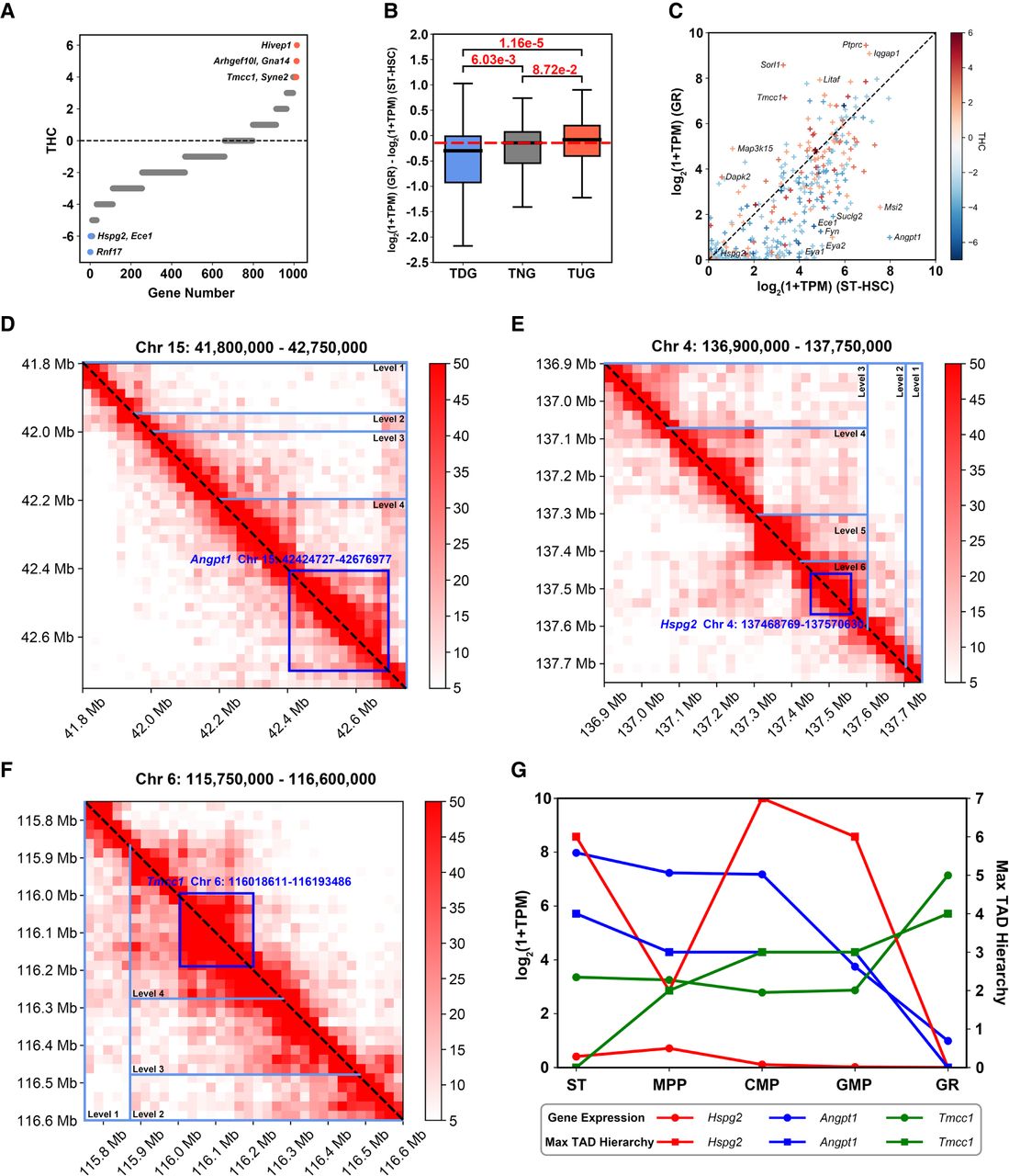
THC unveils genomic dynamics during cellular differentiation. (A) Distribution of THCs of 1013 eligible genes. Genes are sorted in ascending order according to their THCs, for genes with the same THC, we sorted them by string order of their gene names. (B) Boxplot illustrating gene expression variations for TDGs, TNGs, and TNGs between ST and GR (P-values: Wilcoxon rank-sum test). (C) Scatter plot showing gene expression of TUGs and TDGs in GR and ST, with color representing THCs (from low to high, ranging from blue to red). Some representative genes are labeled with their gene symbols. Hi-C submatrices near Angpt1 (D), Hspg2 (E), and Tmcc1 (F), with GR displayed below the diagonal and ST above. Dark blue boxes indicate the locations of genes, and light blue triangles represent hierarchical TADs. (G) Expression of Angpt1, Hspg2, and Tmcc1, along with their corresponding max TAD hierarchies in each cell type (max TAD hierarchy of a gene is defined as the largest level of all TADs containing that gene, and we say that max TAD hierarchy of a gene is zero if there is no TAD containing it) in the GR path.


