
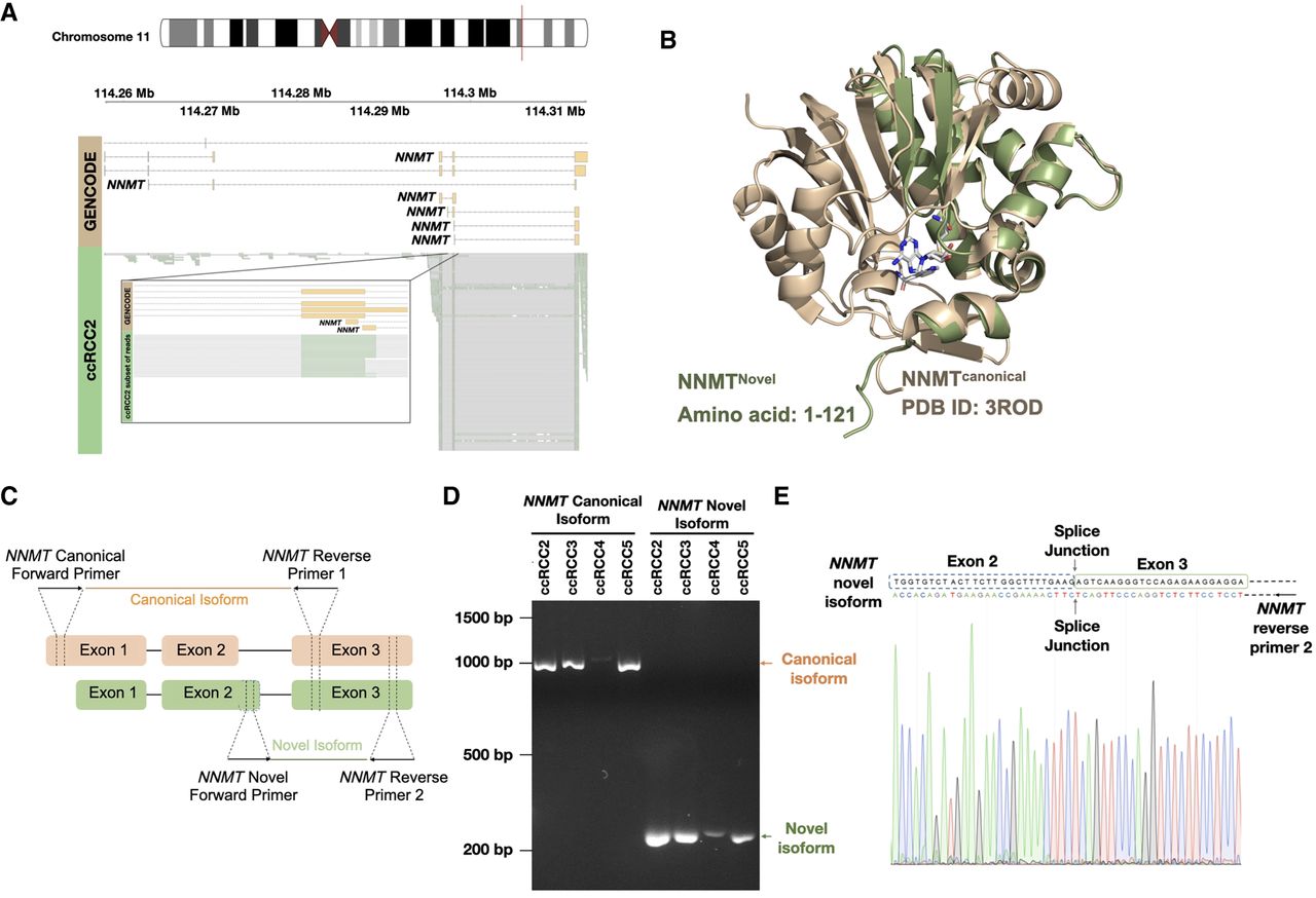
NNMT novel transcripts. (A) Reads that align with the NNMT novel transcript. (B) Alphafold3 structure of the NNMT novel isoform aligned to the canonical isoform of NNMT (PDB ID: 3ROD). (C) Representation of the primer design strategy to validate and sequence the novel and canonical transcripts of NNMT. In the novel isoform, the dotted box indicates the position of the unique sequence at the 3′ end of exon 2. (D) Agarose gel (2%) electrophoresis image of PCR validation of different NNMT transcripts. All lanes are marked with corresponding tumor samples and product names. The canonical transcript is amplified with canonical forward primer and reverse primer 1 (see orange arrow). The novel transcript is amplified with a novel forward primer and reverse primer 2 (see green arrow). (E) Sanger sequencing result of NNMT novel transcript. Reverse sequencing confirmed the unique sequence at the novel transcript's 3′ end of exon 2. The splice junction between exon 2 and exon 3 is marked.


