
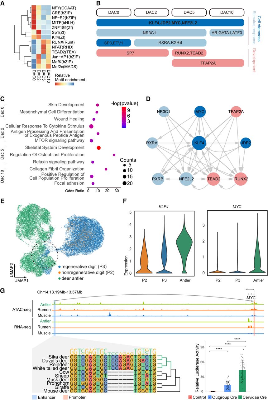
Hub TFs in antler regeneration. (A) Hierarchical clustering heatmap of the top 5 enriched motifs for each stage in antler regeneration. (B) Function and time course schema of top 10 TFs for each stage, suggesting four core TFs and the dynamics from cell stemness and stimulus response to development. (C) Functional enrichment of shared target genes of hub TFs in each antler regeneration stage. (D) Hierarchical structure of 10 hub TFs subnetwork in antler regeneration at dac5 with a similar pattern with time course schema. (E) UMAP projection of PMC cell lineage from antler and mouse digit tips distinguishes PMC of antler and mouse. Dash circle highlights the shared activated stem cell in antler and regenerative digit tip. (F) Higher KLF4 and MYC expression in PMC of deer antler compared with mouse digit tips. (G) Genome track of an antler-specific element near MYC (top), sequence alignment (bottom left), and luciferase assay (bottom right) suggest cervid-specific insertion has significantly increased the expression of MYC in the antler.


