
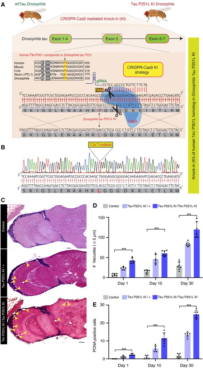
CRISPR-Cas9-mediated knock-in model of frontotemporal dementia in Drosophila. (A) CRISPR-Cas9 gene editing strategy to knock-in the human TAU P301L homologous mutation in Drosophila, Tau P251L, located in exon 5 of Drosophila tau. (B) Successful mutation in homozygous Tau P251L knock-in flies. (C,D) Hematoxylin and eosin staining reveals evidence of neurodegeneration as seen by an increased number of brain vacuoles (arrowheads) with age in homozygous and heterozygous knock-in animals. (C) Scale bar represents 10 µm. (E) Neurodegeneration is accompanied by abnormal cell-cycle reentry as marked by proliferating cell nuclear antigen (PCNA) staining. Flies are 30 d old in C and the age indicated in the figure labels in D,E. (D,E) n = 6 per genotype and time point. Data are presented as mean ± SD. (***) P < 0.001, one-way ANOVA with Tukey post-hoc analysis.


