
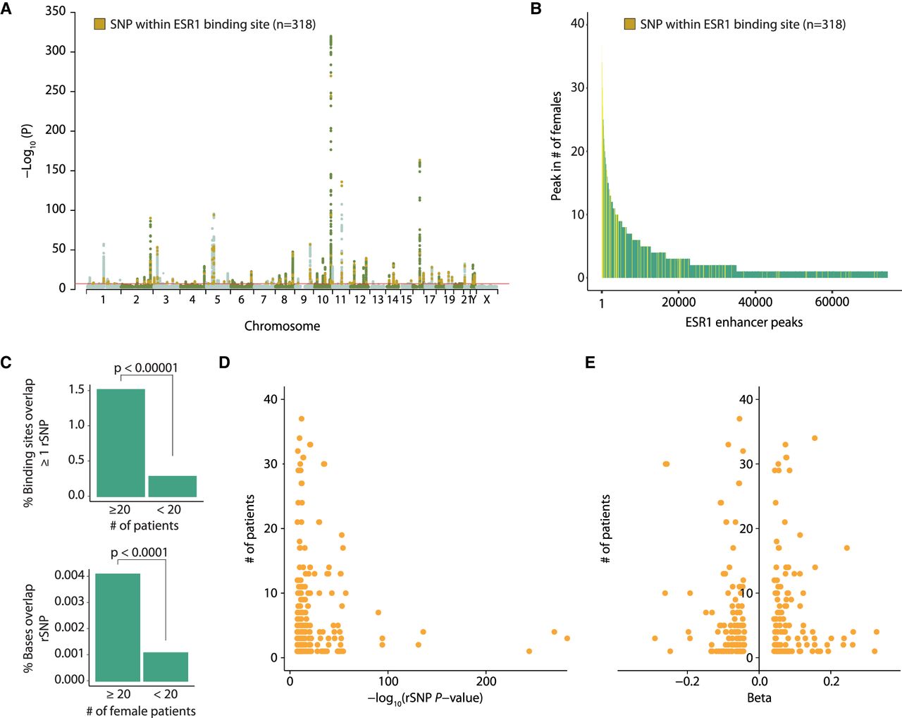
ESR1+ breast cancer rSNPs are enriched at regions with low inter-patient heterogeneity in ESR1. (A) Manhattan plot of ESR1+ breast cancer risk SNPs (rSNPs) with genome-wide significance originating from Michailidou et al. (2017). Highlighted in orange are 318 rSNPs, for which the coordinates intersect with one of the 74,438 ESR1 peaks found among 40 female breast cancer patients. (B) The position of these 318 rSNPs in the ranked peaks introduced in Figure 2A. (C, top) Comparison (Fisher's exact test) of the percentage of ESR1 peaks with which coordinates overlap with at least one rSNP coordinate, for common and less common ESR1 peaks. (Bottom) Comparison (Fisher's exact test) of the percentage of bases, present in common or less common ESR1 peaks, that overlap with at least one rSNP coordinate. (D) Correlation between the P-value of rSNP (x-axis) and its position in the ranking of ESR1 peaks introduced in Figure 2A (y-axis). If multiple rSNPs overlapped the same ESR1 peak, the strongest P-value was used for analysis. (E) Overview of beta values corresponding to rSNPs with which a coordinate intersected an ERE. Negative beta values correspond with rSNPs that confer less risk to breast cancer, whereas positive beta values correspond to increased risk of ESR1+ breast cancer.


