
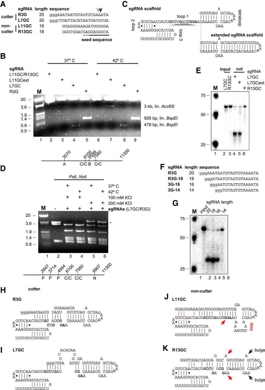
Sequence-dependent in vitro cleavage of oligonucleotides and plasmid DNA by the sgRNA/Cas9 complex. (A) Sequences of sgRNAs with observed cleavage sites indicated by arrow heads. Small letter guanosines used for in vitro transcription are not present in the target DNA sequence. The seed sequence is indicated by a line at the bottom. (B) Agarose gel showing Cas9-mediated cleavage of the 11.3 kb Dscam 3–5 plasmid for 24 h with indicated sgRNAs. Plasmids were cut with either Acc65I (lanes 2, 3, 6, and 7) or BspEI (lanes 4, 5, 8, and 9) after Cas9 cleavage. The line at the bottom shows a map of the plasmid with restriction sites indicated. Size markers are EcoRI/HindIII digested λ DNA of 20 kb, 3.6 kb, 1.9 kb, and 0.8 kb. (C) Structure of the sgRNA scaffold from cocrystallization with Cas9 (Nishimasu et al. 2014). Vertical or horizontal lines indicate Watson–Crick base-pairing, and dots or dashed lines indicate non-Watson–Crick base-pairing. Nucleotides base-pairing in loop 1 are bold. Additional base-pairing found in the tracrRNA–crRNA heterodimer is indicated in the extended scaffold (Jinek et al. 2012). (D) Agarose gel showing Cas9-mediated cleavage of the 11.3 kb Dscam 3–5 plasmid for 24 h with indicated sgRNAs L7GC and R3G. Plasmids were cut with PstI and NotI. The star denotes incomplete cleavage by NotI and the line at the bottom shows a map of the plasmid with restriction sites indicated. Size markers are EcoRI/HindIII digested λ DNA of 20 kb, 3.6 kb, 1.9 kb, and 0.8 kb. (E) Denaturing acrylamide gel showing Cas9-mediated cleavage of synthetic oligonucleotides with indicated sgRNAs. (F) Sequences of sgRNAs with variable length. Small letter guanosines used for in vitro transcription are not present in the target DNA sequence. (G) Denaturing acrylamide gel showing Cas9-mediated cleavage for 1 h of synthetic oligonucleotides with indicated sgRNAs of variable length. (H–K) Structure of sgRNAs. Nucleotides base-pairing in loop 1 are bold. Red lines in J and K indicate potential base-pairing with nucleotides in loop 2. The red arrow in J indicates the sequence complementarity leading to a bulge in the tetraloop. The red arrows in K indicate a duplication of the bulge structure present in the tetraloop.


