
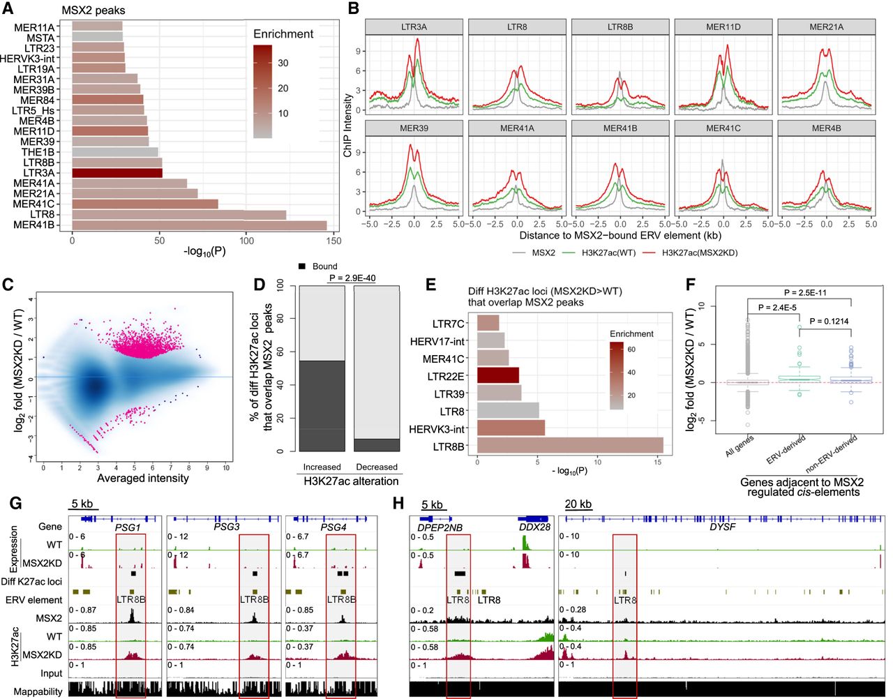
MSX2 restricts the expression of some STB genes in human TSCs through the binding of ERV-derived enhancers. (A) Enrichment of ERV families within MSX2 peaks. The top 20 families as ranked by P-values are presented. (B) Averaged curves show the ChIP intensity of MSX2 and H3K27ac (WT vs. MSX2KD) flanking the MSX2-bound elements from the top ten ERV families. (C) MA plot shows the differential H3K27ac peaks between WT and MSX2KD human TSCs. (D) Comparison of the MSX2-bound percentages between genomic loci with increased or decreased H3K27ac levels after MSX2KD. P-value calculated with Fisher's exact test is denoted. (E) Enrichment of ERV families within MSX2-bound loci that have increased H3K27ac levels after MSX2KD. (F) Comparison of the altered expression (MSX2KD vs. WT) across different groups of genes defined based on their association with MSX2-regulated enhancers. A threshold of <10 kb from TSSs was used to group genes. MSX2-regulated enhancers are further classified as two groups (ERV-derived or non-ERV-derived) based on their overlapping with ERV elements. P-values calculated from two-sided student's t-test are denoted. (G,H) Representative IGV tracks showing several LTR8B- or LTR8-derived enhancers that are likely mediating the MSX2-dependent repression of STB genes, including PSG1/3/4, DPEP2NB, DDX28, and DYSF. The RNA-seq and H3K27ac ChIP-seq data in WT and MSX2KD TSCs were adopted from Hornbachner et al. (2021).


