
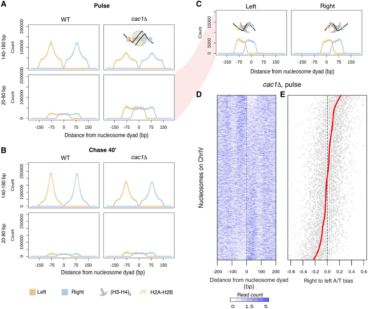
Subnucleosomal fragments captured in cac1Δ cells reveal nucleosome assembly intermediates. (A,B) Aggregate signals of the left (orange) and right (blue) ends of fragments surrounding the dyads of high-confidence nucleosomes in nascent chromatin (A) and mature chromatin (B) in the two strains. Fragments are divided into nucleosome-sized fragments (140–180 bp in length; top panels) and subnucleosomal-sized fragments (20–80 bp in length; bottom panels). A diagram depicting the fragment ends in relation to the nucleosome is inset in the panel. (C) Fragment ends of the subnucleosomal fragments in the nascent chromatin of cac1Δ cells are plotted separately by the location of the fragment midpoint relative to the nucleosome dyads. (D) Heatmap depicting occupancy of subnucleosomal fragments surrounding individual nucleosomes. Each row corresponds to a high-confidence nucleosome on Chr IV. Rows are ordered by increasing right-to-left occupancy bias (calculated as the log2 ratio) of the subnucleosomal fragments. (E) Dot plot showing the right-to-left A/T bias of DNA sequence associated with individual nucleosomes in the same order as in D. The red line denotes the fitted smooth spline curve.


