
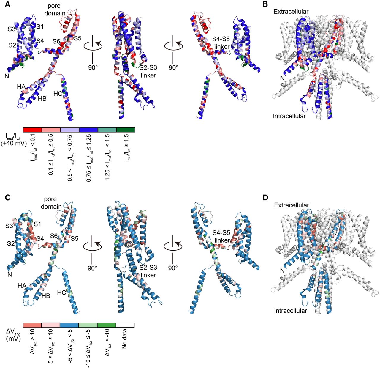
Structural basis of KCNQ4 missense SNVs. (A) Cartoon diagrams showing three different views of a KCNQ4 monomer denoting specific domains. Currents recorded at +40 mV were mapped onto the monomer structure (covering residues S74-Y367 and D524-G588). Variants were colored based on currents normalized to WT. The mean current of each amino acid is calculated by averaging the current values of all four to seven missense SNVs at this site. N-terminal front (M1-S73), S3–S4 linker (T193-A199), helix A–helix B linker (Y368-V523), and C-terminal end (R589-D695) residues are not available in the cryo-EM structure. Colors indicating the currents were drawn using PyMOL. (B) Normalized currents were mapped onto one subunit of the tetramer structure. (C) KCNQ4 protein monomer residues were colored by the shifts in activation V1/2. White indicates that the currents of all four to seven missense SNVs at this site are small, and the V1/2 cannot be determined. (D) Activation V1/2 was mapped onto one subunit of the tetramer structure.


