
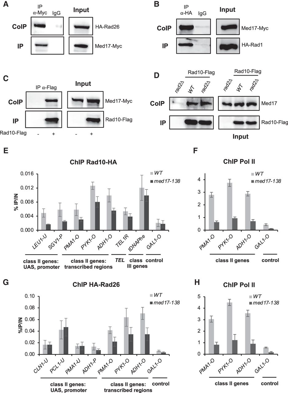
Rad1, Rad10, and Rad26 coimmunoprecipitate with Mediator, and the loss of Mediator head in the med17-138 mutant decreases Rad10 chromatin binding. (A–D) Co-IP between Rad1, Rad10, and Rad26 and Mediator. Inputs are shown in right panels. (A) Mediator was immunoprecipitated from crude yeast extracts via Med17-Myc subunit with α-Myc antibody (IP), and western blotting with α-HA antibody detected HA-Rad26 (Co-IP). (B–D) HA-Rad1 (B) or Rad10-Flag (C,D) was immunoprecipitated with α-HA or α-Flag antibody, respectively, and analyzed by western blotting with α-Myc antibody (B,C) against Med17-Myc subunit or rabbit polyclonal α-Med17 antibody (D) to detect the Med17 (Mediator) subunit (Co-IP) (left panels). WT (A–D) or rad2Δ (D) strains were used. IgG indicates a control immunoprecipitation with IgG magnetic beads only (A,B). A strain carrying nontagged Rad10 was used as a negative control in C and D. (E–H) Effect of the med17-138 mutation on Rad10, Rad26, and Pol II occupancies at selected regions. Quantitative ChIP assays were performed using α-Rpb1 antibody (Pol II) (F,H), and α-HA antibody against Rad10-HA (E) or HA-Rad26 (G). Cells were grown in selective SD medium complemented with amino acids at 25°C and then shifted for 45 min at 37°C. GAL1-O amplicon was used as a negative control. Quantities were normalized to qPCR performed on input DNA and are expressed as a percentage. The indicated value is the mean of three biological replicates, and error bars represent the standard deviation.


