
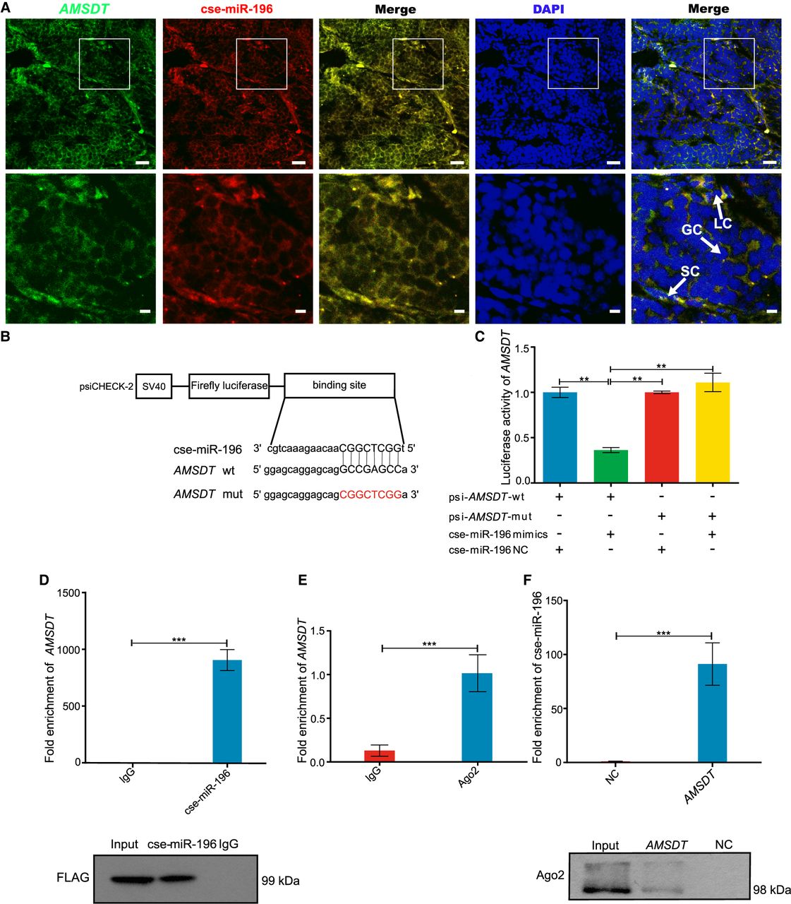
AMSDT physically associates with cse-miR-196. (A) Representative images of confocal micrographs of the subcellular localization and expression of AMSDT (green) and cse-miR-196 (red) in tongue sole testis. Nuclei were counterstained with DAPI (blue). The bottom row shows enlargement of the regions outlined in the top row. Scale bars in top rows are 20 μm, and bars in bottom rows are 5 μm. (B) Putative binding sites of cse-miR-196 on AMSDT. (C) Luciferase activity of AMSDT in HEK293T cells transfected with cse-miR-196 mimics. Luciferase activity was normalized to Renilla luciferase activity of AMSDT. (D) Ago2 immunoprecipitation executed in HEK293T cells that are stably expressing Ago2 (tongue sole), AMSDT, and cse-miR-196, followed by western blot and qRT-PCR to detect AMSDT (top) and Ago2 protein (bottom), respectively. (E) Ago2 immunoprecipitation was executed in tongue sole testis, followed by qRT-PCR to detect AMSDT. (F) RNA pull-down of tongue sole testis lysates, followed by qRT-PCR and western blot to detect the enrichment of cse-miR-196 and Ago2 protein. The NC and AMSDT in the figure represent the AMSDT-mut and AMSDT-wt probe captured fraction group, respectively. The bars in C–F represent triplicate mean ± SD values from three biological replicates. (n = 3.) (∗∗) P < 0.01, (∗∗∗) P < 0.001, two-tailed t-test.


