
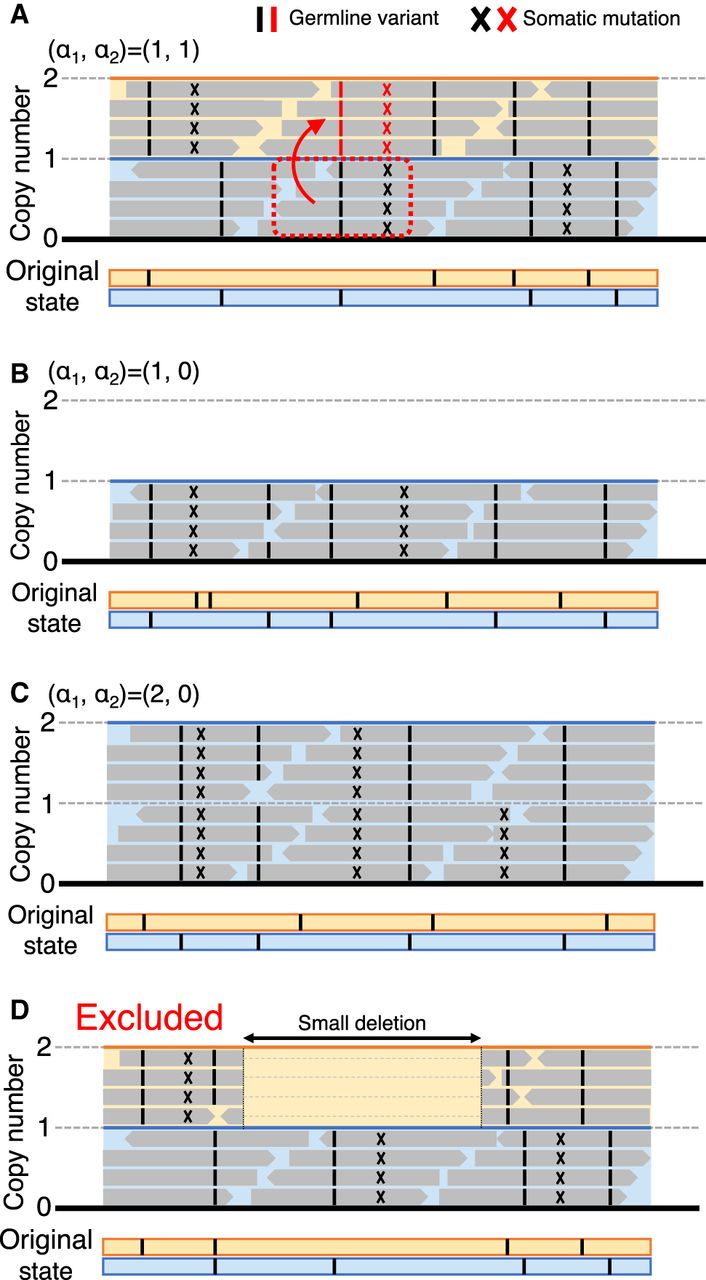
Illustrations of short-read sequencing data mapped on the reference sequence. The original state before somatic mutations occur is presented at the bottom using the blue and orange lines, representing the paternal and maternal chromosomes, where only germline variants are present (black bars). Somatic mutations can be detected in the short-read data, presented by X. (A) A hypothetical case of (α1, α2) = (1, 1), where a gene conversion event from the paternal to the maternal chromosomes occurred (boxed by the red dotted line). The mutated sites transferred by gene conversion are shown in red. (B,C) Cases of (α1, α2) = (1, 0) and (2, 0). (D) Cases with a small deletion within a region of (1, 1) that potentially causes false evidence for gene conversion (excluded from the analyses).


