
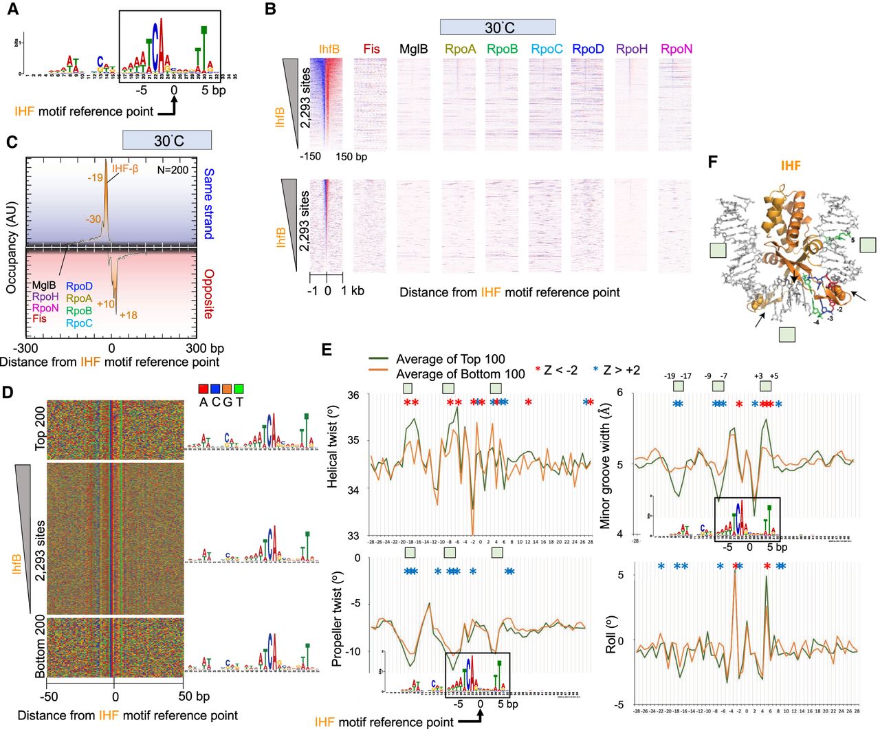
Figure 6.
Genome-wide interactions of IHF. (A) MEME motif derived from the top 500 IHF-bound locations as described in Figure 2A. (B) Heatmap occupancy of indicated protein targets at 30°C distributed around the IHF motif reference point. For plotting details, see Figure 2B. (C–E) Distribution of IHF and other target occupancies at 30°C (C), nucleotide sequence (D), and DNA shape around IHF motifs (E). For plotting details, see Figure 2, D through F. Green boxes correspond to regions shown in panel F. (F) Crystal structure of IHF (Rice et al. 1996). Select motif base coloring is as defined in panel D.


