
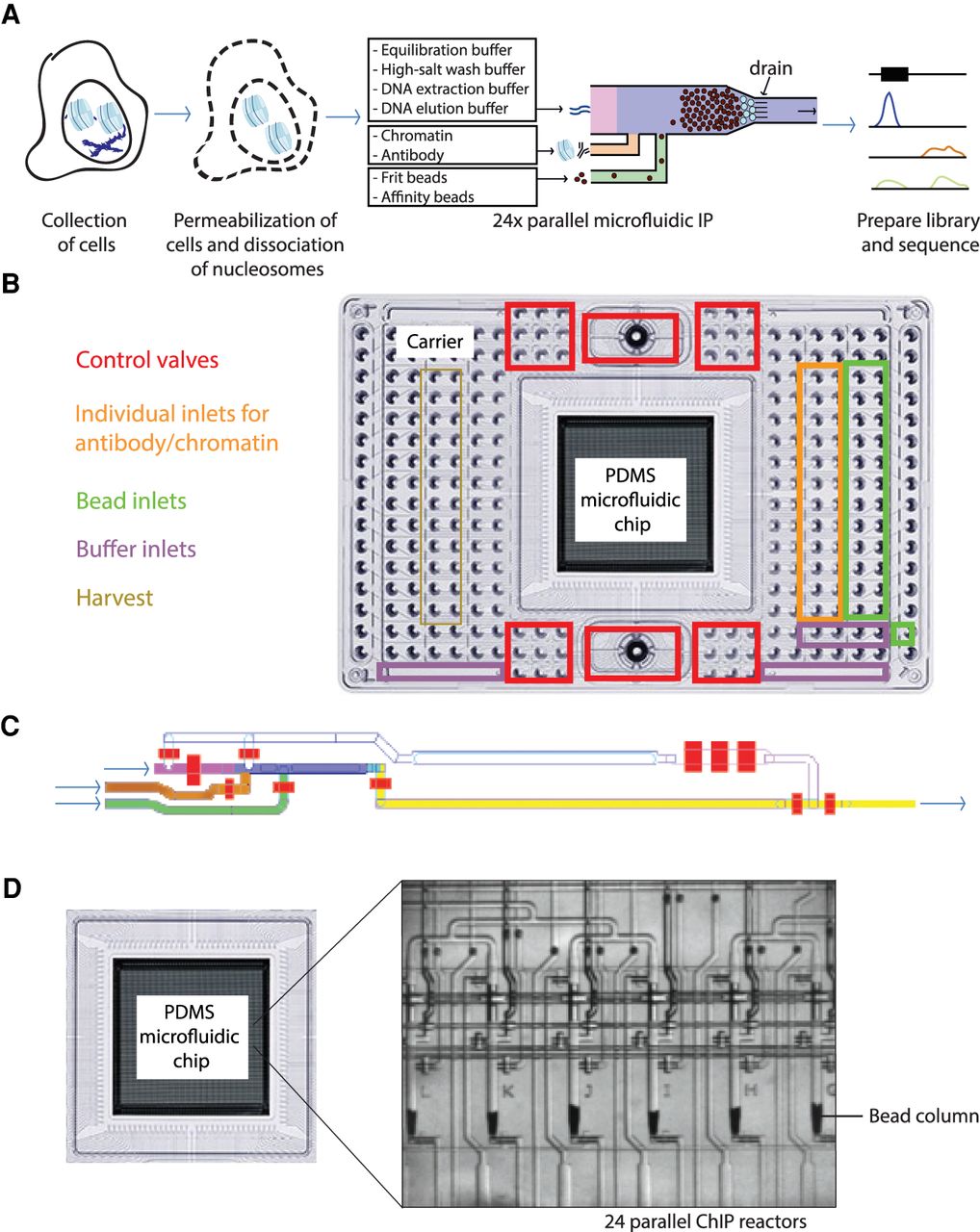
Overview of the microfluidic chip design for automated microfluidic chromatin immunoprecipitation. (A) Workflow of automated microfluidic ChIP-seq. (B) Overview of the interface plate. At the sides are the inlets, whereas the PDMS microfluidic chip containing the microreactors is located in the center. (C) Architecture of PDMS microfluidic chip, also referred to as integrated fluidic circuit (IFC). The bead inlet is in green; the antibody and chromatin inlets, orange; the channel in which the bead column is constructed, blue; the inlet for various buffers needed in the workflow, pink; and the waste and harvest outlet, yellow. The control valves are colored red. (D) Phase contrast image of six out of 24 parallel microfluidic bead columns on every chip.


