
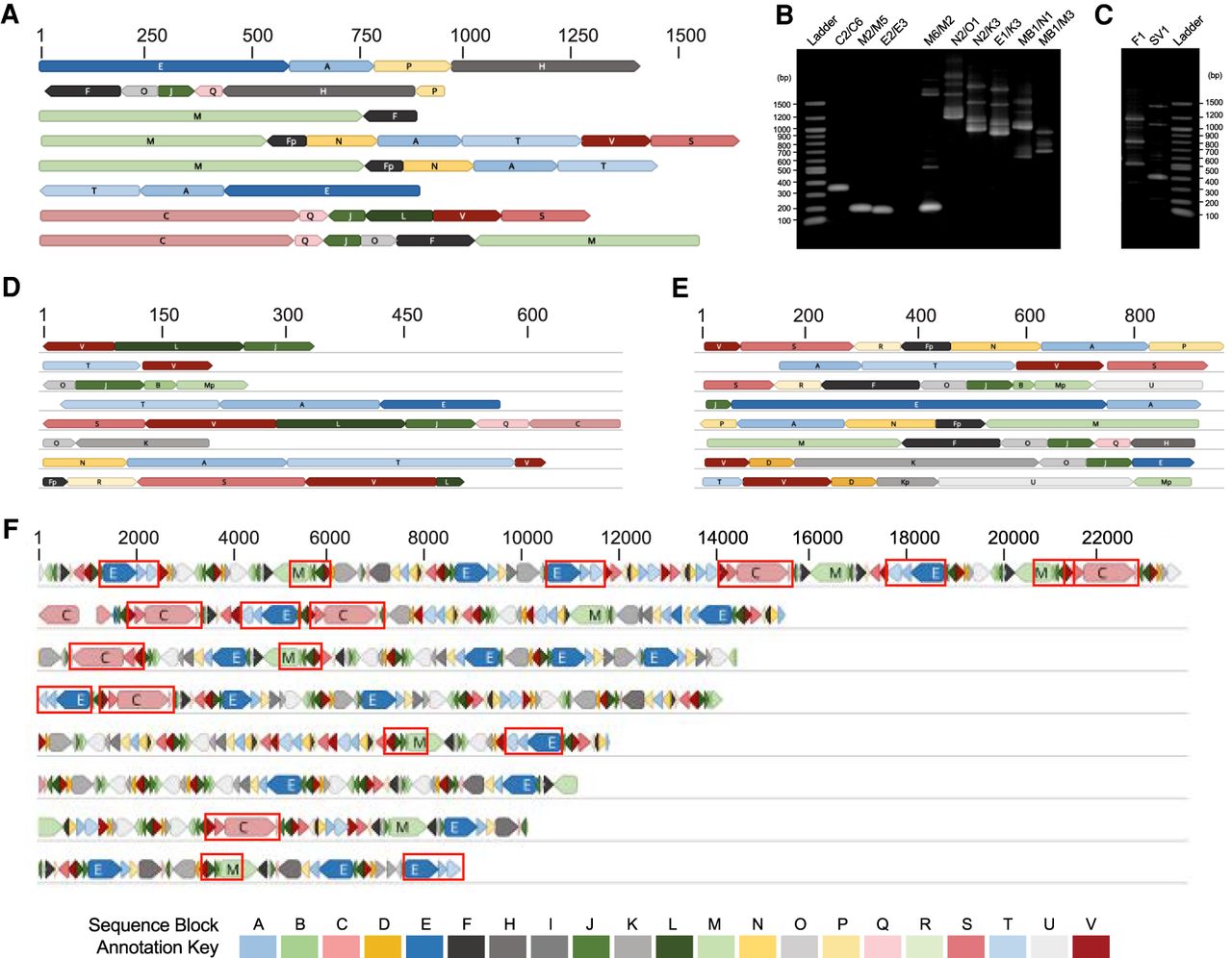
Sequence evidence of fragmented mtDNA and full-length protein-encoding genes. (A, D–F) Each row of colored blocks represents individual, annotated T. gondii (A) mtDNA-specific PCR amplicons; (D) EST reads; (E) Sanger genomic reads; or (F) Nanopore reads. Sections of reads containing identical sequence to other mtDNA reads are called “sequence blocks” (SBs) and are colored and labeled with a unique color and letter corresponding to the key, Table 1 and Figure 2. Shades of blue, red, and green represent different SBs found in cob, coxI, and coxIII, respectively. Orientation of a block is indicated by the point on each block. Red boxes in F indicate complete protein-encoding genes. SBs located on the ends of reads may be incomplete. (B) Genomic DNA from mitochondrial-enriched fractions assayed with different primer pairs (Supplemental Table S1) as indicated. Lanes 2–4 represent a small fragment of each cytochrome gene. (C) Single primers produce multiple amplicons. Scale (in bp) is as indicated in each panel. Additional reads are in Supplemental Figures S1–S4.


