
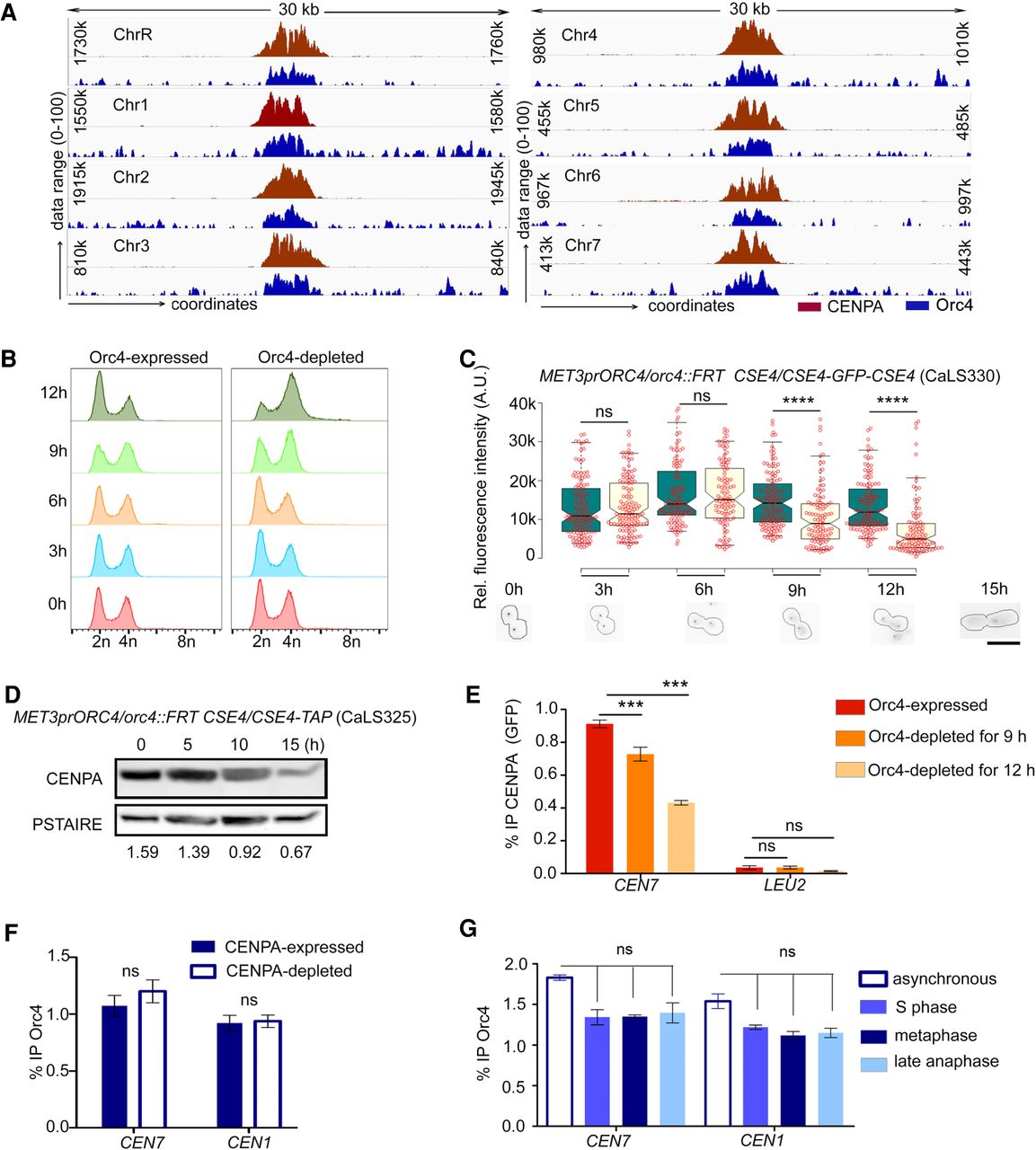
Centromeric localization of Orc4 stabilizes CENPA. (A) A 30-kb region harboring each centromere (x-axis) was plotted against the subtracted ChIP sequencing reads (y-axis) for CENPA (red) and Orc4 (blue). (B) Flow cytometric analyses of orc4 mutants in Orc4-expressed versus Orc4-depleted conditions at the indicated time of incubation. (C) Relative fluorescence intensities of CENPA (GFP) clusters in orc4 mutant CaLS330 grown at indicated time points in permissive (green) and nonpermissive (yellow) conditions show a significant reduction of GFP upon depletion of Orc4. Scale bar, 10 µm. t-test; (****) P-value < 0.0001, (ns) P-value > 0.05; n ≥ 100. (D) Western blot of the whole-cell lysate of CaLS325 (METprORC4/orc4::FRT CSE4/CSE4-TAP) using anti–Protein A antibodies showed a time-dependent decrease in CENPA levels upon Orc4 depletion when normalized with the loading control, PSTAIRE. (E) ChIP-qPCR using anti-GFP (CENP-A) antibodies revealed reduced CENPA enrichment at the centromere upon Orc4 depletion in CaLS330 when grown in nonpermissive media for 9 and 12 h. Two-way ANOVA; (***) P-value < 0.001; (ns) P-value > 0.05; n = 3. (F) Orc4 ChIP-qPCR revealed no significant reduction in centromeric Orc4 when CENPA was depleted for 8 h in YP with dextrose in the strain CAKS3b (cse4/PCK1prCSE4) (Sanyal and Carbon 2002). Two-way ANOVA; (ns) P-value > 0.05; n = 3. (G) ChIP-qPCR revealed the Orc4 enrichment at CEN7 in various stages of the cell cycle: hydroxyurea treated (S phase), nocodazole treated (metaphase), cdc15 mutant (late anaphase). Percentage IP values for Orc4 ChIP at CEN7 were normalized with noncentromeric regions enriched with Orc4. One-way ANOVA; (***) P-value < 0.001; (ns) P-value > 0.05; n = 3.


